Key Concepts

Chapter 1 Key Concepts


Please go to expertconsult.com for supplemental chapter material.


The electrocardiogram (ECG or EKG) is a special graph that represents the electrical activity of the heart from one instant to the next. Thus, the ECG provides a time-voltage chart of the heartbeat. For many patients, this test is a key component of clinical diagnosis and management in both inpatient and outpatient settings.


The device used to obtain and display the conventional ECG is called the electrocardiograph, or ECG machine. It records cardiac electrical currents (voltages or potentials) by means of conductive electrodes selectively positioned on the surface of the body.


For the standard ECG recording, electrodes are placed on the arms, legs, and chest wall (precordium). In certain settings (emergency departments, cardiac and intensive care units [CCUs and ICUs], and ambulatory monitoring), only one or two “rhythm strip” leads may be recorded, usually by means of a few chest electrodes.


Essential Cardiac Electrophysiology


Before basic ECG patterns are discussed, we will review a few simple principles of the heart’s electrical properties.


The central function of the heart is to contract rhythmically and pump blood to the lungs for oxygenation and then to pump this oxygen-enriched blood into the general (systemic) circulation.


The signal for cardiac contraction is the spread of electrical currents through the heart muscle. These currents are produced both by pacemaker cells and specialized conduction tissue within the heart and by the working heart muscle itself.


Pacemaker cells are like tiny clocks (technically called oscillators) that repetitively generate electrical stimuli. The other heart cells, both specialized conduction tissue and working heart muscle, are like cables that transmit these electrical signals.


Electrical Activation of the Heart


In simplest terms, therefore, the heart can be thought of as an electrically timed pump. The electrical “wiring” is outlined in Figure 1-1.


Normally, the signal for heartbeat initiation starts in the sinus or sinoatrial (SA) node. This node is located in the right atrium near the opening of the superior vena cava. The SA node is a small collection of specialized cells capable of automatically generating an electrical stimulus (spark-like signal) and functions as the normal pacemaker of the heart. From the sinus node, this stimulus spreads first through the right atrium and then into the left atrium.


Electrical stimulation of the right and left atria signals the atria to contract and pump blood simultaneously through the tricuspid and mitral valves into the right and left ventricles. The electrical stimulus then reaches specialized conduction tissues in the atrioventricular (AV) junction.


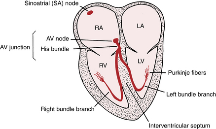
The AV junction, which acts as an electrical “relay” connecting the atria and ventricles, is located at the base of the interatrial septum and extends into the interventricular septum (see Fig. 1-1).

Only gold members can continue reading. Log In or Register to continue

Stay updated, free articles. Join our Telegram channel

Jun 11, 2016 | Posted by in CARDIOLOGY | Comments Off on Key Concepts

Full access? Get Clinical Tree

Get Clinical Tree app for offline access