Figure 12.1
Apical two chamber view of a patient with inferior and inferobasal scarring (arrows) after myocardial infarction
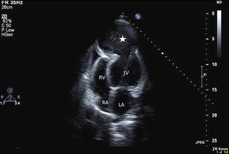
Figure 12.2
Apical four chamber view showing massive apical aneurysm (star)

Figure 12.3
Modified parasternal long axis view showing ruptured posterior papillary muscle (arrows)
ECG tracings are analyzed
Assessment wall motion and contractility changes helps evaluate patients for presence of obstructive coronary artery disease. Degree of mitral regurgitation as well as changes in RVSP may be evaluated.
Assessment for ischemic mitral regurgitation
Stress test may also be ordered in order to evaluate valvular disease
Pharmacological Stress Modalities (Video 12.3)
Used for patients who are unable to exercise or have low exercise capacity
dobutamine echocardiography is commonly used< div class='tao-gold-member'>Only gold members can continue reading. Log In or Register to continue
Stay updated, free articles. Join our Telegram channel

Full access? Get Clinical Tree


