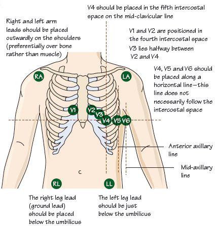
Fig. 1.2 ECG lead placement for an exercise ECG – in a resting ECG the leads to the legs are attached to electrodes just above the ankles. The ECG can be extended further beyond V6, to include leads V7–9, which extend posteriorly on the left chest. The leads can also be extended further rightward beyond lead V1, as ‘right-sided chest leads’.

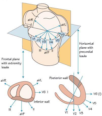
Fig. 1.3 The direction from which the basic 12-leads of the ECG examine the heart.

The electrocardiogram (ECG) is a wonderful tool, cheap, widely available, and incredibly useful. It informs diagnosis, guides and assesses the response to therapy and provides vital data on prognosis. In epidemiological use it gives great insights, e.g. it informs us that 30% of myocardial infarctions (MIs) are clinically silent, and that hypertensive heart disease when associated with certain ECG changes has a high mortality. The ECG informs us not only in acquired heart disease but also in genetic disease, e.g. hereditary long QT or Brugada syndrome. The diagnostic role extends beyond cardiac disease to pulmonary emboli, electrolyte imbalance, rheumatic disease, fitness level, liver disease, diabetes, starvation, etc. It is probably the most useful investigative tool in the whole of medicine.
A brief history of the ECG
Stay updated, free articles. Join our Telegram channel

Full access? Get Clinical Tree


