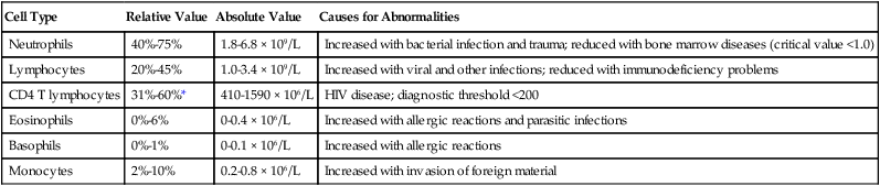
Interpreting Clinical and Laboratory Data


Interpreting Clinical and Laboratory Data


Richard H. Kallet


Interpreting Clinical Laboratory Tests


This chapter primarily discusses common blood tests performed on patients admitted to the hospital. These tests are done to evaluate the general health status of the patient, identify organ system dysfunction, detect the presence of infection, and determine the effects of therapy. The respiratory therapist (RT) must be familiar with these tests to understand the overall clinical status of patients under their care. The RT must be able to recognize how some abnormalities influence pulmonary function specifically. Sometimes the RT must alter his or her approach to practice based on abnormal laboratory test results.


This chapter also presents a brief review of fundamental physiologic concepts related to these tests. Comprehensive tables provide detailed information that can be used as a quick reference for each test. The chapter text provides a more general explanation on the significance of these tests and how they fit into an overall assessment of a patient’s status.


Introduction to Laboratory Medicine


Laboratory medicine involves the study of patient tissue and fluid specimens. It is divided into five major disciplines. Clinical biochemistry involves the analysis of blood, urine, and other bodily fluids primarily for electrolytes and proteins; hematology analyzes the cellular components of blood. The analysis of blood and other bodily fluids for the presence of infectious agents is the purview of clinical microbiology; this includes the subspecialties of identifying bacteria (bacteriology), viruses (virology), fungi (mycology), and parasites (parasitology). A closely related discipline involves the analysis of the immune system (immunology) focusing on autoimmune and immunodeficiency diseases. Finally, the analysis of tissue for diagnosing diseases is the purview of the anatomic pathology service.


Reference Range


Laboratory tests are employed to determine a patient’s health status and aid medical decisions. It is important to determine whether a specific test result falls within an expected range of values considered to be “normal.” The notion of normal is problematic, however. Early on in the history of laboratory medicine, tests to determine the normal range for blood chemistry and hematology were done primarily on small convenience samples of subjects who were not representative of the larger population in terms of age, gender, race, and ethnicity. An additional problem is that the term normal is not the same as healthy. The best example is cholesterol. A normal range of cholesterol found in most Americans puts them at risk of cardiovascular disease and cannot be considered healthy.


Beginning in the 1970s,1 the term normal ranges was slowly replaced with more appropriate terms such as reference ranges, biologic reference intervals, and expected value.2 This change in terminology acknowledged that what we consider normal must take into account variations related to age, gender, race, and ethnicity, which change over time as the demographic composition of society changes. A reference range sets the boundaries for any analyte (e.g., electrolyte, blood cell, protein, enzyme) that would likely be encountered in healthy subjects. This range would encompass the variability reflected in the larger, presumably healthy population.


Reference ranges vary from laboratory to laboratory for various reasons, including differences in measurement techniques, the populations of healthy individuals used to establish the reference intervals, or analytic imprecision when the intervals were constructed. Most differences in reference ranges are relatively small, with reasonably close agreement between most laboratories.2 The reference ranges and critical values given in this chapter are from a single institution, and they serve as representative examples. RTs must become familiar with the reference ranges used at their institutions.


Critical Test Value


A critical test value is a result significantly outside the reference range and represents a pathophysiologic condition. A critical value may be potentially life-threatening unless corrective action is taken promptly. Critical values are reported in the hospital to alert caregivers as well to decrease medical errors and protect patients.


Typically, critical values are communicated by telephone from the clinical laboratory to the general ward or intensive care unit where the patient is situated. The nurse or RT who receives these results is required to read-back the critical value to the clinical laboratory. This requirement is to ensure that the correct information has been communicated. It is the responsibility of the nurse or RT to communicate the critical value in a timely fashion to the physician caring for the patient. The same read-back procedure is used. All communication of critical test values is documented in the medical record.


In this chapter, critical values are listed along with common pathophysiologic states with which they commonly occur. Not all clinical analytes have an associated critical value. For some tests, there is no general agreement on what a critical value would be. Others have only a one-sided value that exists below or above a critical threshold; this is true particularly for substances that do not normally appear in the blood. For example, certain enzymes and proteins are released only after extensive cellular damage following injury (see later section on enzyme tests). Under normal circumstances, these proteins or enzymes may be virtually undetectable in the serum or plasma.


When interpreting derangements for any test result, the clinician must consider the context of the change. In a patient with chronic renal disease, a serum creatinine of 3.0 mg/dl (approximately twice the upper limit of normal) would not be considered urgent. However, in a patient who presents with a bloodstream infection (i.e., sepsis) and hypotension, a sudden increase in serum creatinine to 3.0 mg/dl would be considered critical because it indicates acute kidney injury in the context of rapidly developing clinical instability.


Complete Blood Count


The complete blood count (CBC) provides a detailed description of the number of circulating white blood cells (WBCs), called leukocytes; red blood cells (RBCs), called erythrocytes; and platelets, called thrombocytes. The WBC count is made up of five different types of cells and is reported under the differential. RBCs are evaluated for size and hemoglobin content. The platelets are evaluated for number present. Table 16-1 lists the normal CBC results for adults.


Elevation of the WBC count is termed leukocytosis. It results from numerous problems, including stress, infection, and trauma. The degree of leukocytosis reflects the severity of infection. A significantly elevated WBC count (>20 × 103/mcl) suggests the presence of a serious infection and that the patient’s immune system is generating a significant response. In contrast, leukopenia (or leukocytopenia) is a WBC count below normal that often occurs when the patient’s immune system is overwhelmed by infection. Other important causes of leukopenia include bone marrow diseases (e.g., leukemia, lymphoma), influenza, systemic lupus erythematosus, tuberculosis, and acquired immunodeficiency syndrome (AIDS). Also, chemotherapy and radiation therapy given to cancer patients frequently causes leukopenia.


White Blood Cell Count


White Blood Cell Count Differential

The differential of the WBC count determines the exact number of each type of WBC present in the circulating blood (Table 16-2). Most circulating WBCs are either neutrophils or lymphocytes. Because leukocytosis usually results from only one of the five cell types responding to a problem, significant elevation of the WBC count (>15 × 103/mcl) occurs only when either neutrophils or lymphocytes are responding to an abnormality. Because basophils, eosinophils, and monocytes make up such a small proportion of the circulating WBCs, they are not likely to cause a major increase in the WBC count when responding to disease.


The WBC count differential is best interpreted by determining the absolute count of each WBC; this is calculated by multiplying the percentage of the WBCs under study by the total WBC count. This calculation prevents misinterpretation of the WBC count differential when any one cell type changes in absolute numbers and causes a relative change in the percentage of the other four cell types. For example, if the WBC count doubles because of an increase in neutrophils, the relative value of the other four cells would decrease by half, although their absolute value would not change. Many laboratories report the absolute value for each of the five WBCs to avoid this confusion.


The subanalysis of lymphocytes is important for identifying infection with HIV, the causative agent of AIDS. HIV targets and destroys CD4 T lymphocytes. Opportunistic infections, in particular, Pneumocystis jiroveci pneumonia, generally occur when these lymphocytes decrease to less than 200 × 106/L, and this information is used in making the diagnosis of AIDS.


Elevation of the absolute value of neutrophils is termed neutrophilia. Immature neutrophils are known as bands because of the banded shape of the nucleus. Most bands are located in the bone marrow where they continue to mature. Mature neutrophils are known as segs because of the segmented shape of their nucleus. Severe infection causes the bone marrow to release stores of any available neutrophils, and both bands and segs enter the circulating blood volume. When bands and segs are elevated in the CBC, the patient is likely experiencing a more severe bacterial infection.


A reduced number of circulating neutrophils is termed neutropenia. Although uncommon, neutropenia is characteristic of patients with bone marrow disease (e.g., lymphoma, leukemia), patients undergoing treatment for cancer with chemotherapy or radiation or both, patients with some autoimmune disorders, and HIV-infected patients. Neutropenia puts the patient at risk for the development of infection.


Red Blood Cell Count


The primary function of RBCs or erythrocytes is to supply oxygen to the tissues. The RBC count helps determine the ability of the blood to carry oxygen. An abnormally low RBC count is referred to as anemia and suggests that either RBC production by the bone marrow is inadequate or excessive blood loss has occurred. In either case, the oxygen-carrying capacity of the blood is reduced, and the patient is more likely to experience tissue hypoxia. There are several types of anemia with different causes. Some are related to dietary deficiencies in iron or vitamins (e.g., vitamin B12 and folate). Other causes are related to chronic inflammatory diseases, such as Crohn disease, HIV/AIDS, lymphoma, and autoimmune diseases that result in the destruction of erythrocytes (hemolytic and aplastic anemia). A hereditary cause is sickle cell anemia, which is common in African-Americans. For most forms of anemia, a blood transfusion may be needed if the RBC count is too low. The trigger point for transfusion is based on the hemoglobin or hematocrit measurement rather than the RBC count.


An abnormally elevated RBC count is known as polycythemia. It occurs most often when the bone marrow is stimulated to produce extra RBCs in response to chronically low blood oxygen levels (secondary polycythemia). Polycythemia counteracts the negative side effects of reduced PO2 in the blood by increasing the oxygen-carrying capacity of the blood. Patients who live at a high altitude and patients with chronic lung disease are most likely to experience chronic hypoxia and to develop secondary polycythemia.


In addition to the RBC count, the clinical laboratory reports hemoglobin and hematocrit levels. Hemoglobin is a protein substance with the unique ability to bind with oxygen. Each healthy RBC contains 200 million to 300 million molecules of hemoglobin, for a hemoglobin level of 12 to 17 g/dl in a healthy adult. Patients with an inadequate hemoglobin concentration have reduced oxygen-carrying capacity. In this condition, the RBCs are smaller than normal (microcytic anemia) and lack normal color (hypochromic anemia). The necessity for RBC transfusion depends on the cause of anemia and the patient’s overall condition. Usually a transfusion is triggered by a hemoglobin concentration of approximately 7.0 g/dl or a hematocrit of approximately 21%.

Stay updated, free articles. Join our Telegram channel

Jun 12, 2016 | Posted by in RESPIRATORY | Comments Off on Interpreting Clinical and Laboratory Data

Full access? Get Clinical Tree

Get Clinical Tree app for offline access