Every electrocardiogram (ECG) has nine features that should be examined systematically:
1. Rate and regularity;
2. P-wave morphology;
3. PR interval;
4. QRS complex morphology;
5. ST-segment morphology;
6. T-wave morphology;
7. U-wave morphology;
8. QTc interval; and
9. Rhythm.
Rate, regularity, and rhythm are commonly grouped together. However, to accurately assess rhythm, it is necessary to consider not only rate and regularity but also the various waveforms and intervals.
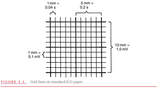
Determination of the ECG features requires understanding of the grid markings provided on the ECG paper (Fig. 3.1). The paper shows thin lines every 1 mm and thick lines every 5 mm. The thin lines, therefore, form small (1 mm) squares and the thick lines form large (5 mm) squares. The horizontal lines facilitate measurements of the various intervals and determination of cardiac rate. At the standard paper speed of 25 mm per second, the thin lines occur at 0.04-second (40-millisecond) intervals and thick lines occur at 0.20-second (200-millisecond) intervals. The vertical lines facilitate measurements of waveform amplitudes. At the standard calibration of 10 mm per mV, the thin lines are at 0.1-mV increments and the thick lines are at 0.5-mV increments. Therefore, each small square is 0.04 second × 0.1 mV, and each large square is 0.20 second × 0.5 mV.
Much of the information provided by the ECG is contained in the morphologies of three principal waveforms: (a) the P wave, (b) the QRS complex, and (c) the T wave. It is helpful to develop a systematic approach to the analysis of these waveforms by considering their:
1. General contours,
2. Durations,
3. Positive and negative amplitudes, and
4. Axes in the frontal and transverse planes.
The guidelines for measuring and estimating these four parameters for each of the three principal ECG waveforms are presented in this chapter. The definitions of the various waveforms and intervals were presented in Chapter 1 in the context of describing ECG recordings of base-to-apex and left- versus right-sided cardiac activity.
The cardiac rhythm is rarely precisely regular. Even when electrical activity is initiated normally in the sinus node, the rate is affected by the autonomic nervous system. When an individual is at rest, minor variations in autonomic balance are produced by the phases of the respiratory cycle. A glance at the sequence of cardiac cycles is sufficient to determine whether the cardiac rate is essentially regular or irregular. Normally, there are equal numbers of P waves and QRS complexes. Either of these may be used to determine cardiac rate and regularity. When in the presence of certain abnormal cardiac rhythms, the numbers of P waves and QRS complexes are not the same. Atrial and ventricular rates and regularities must be determined separately.
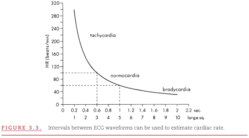
If there is essential regularity in the cardiac rhythm, cardiac rate can easily be determined by counting the number of large squares between cycles. Because each square indicates one fifth of a second and there are 300 fifths of a second in a minute (5 × 60), it is necessary only to determine the number of large squares between consecutive cycles and divide this number by 300. It is most convenient to select the peak of a prominent ECG waveform that occurs on a thick line and then count the number of large squares until the same waveform recurs in the following cycle. When this interval is only one fifth of a second (0.2 second), the cardiac rate is 300 beats per minute; if the interval is two fifths of a second (0.4 second), the cardiac rate is 150 beats per minute; if the interval is three fifths of a second (0.6 second), the cardiac rate is 100 beats per minute, and so forth. Lead II is displayed in Figure 3.2 with the second QRS complex following the onset of the initial QRS complex after four large squares (heart rate = 75 beats per minute).

When the cardiac rate is <100 beats per minute, it is sufficient to consider only the large squares on the ECG paper. When the rate is >100 beats per minute (tachycardia), however, small differences in the observed rate may alter the assessment of the underlying cardiac rhythm, and the number of small squares must also be considered (Fig. 3.3). This illustrates the importance of considering the small squares (0.04 second or 40 milliseconds) rather than the large squares (0.2 second or 200 milliseconds) for estimating rates in the tachycardic range, where small differences in the number of intervals between cardiac cycles result in large differences in the estimated rate. Because there are five small squares in each large square, the number of small squares between successive waveforms of the same type must be divided into 1,500 (6 squares = 250 beats per minute, 7 squares = 214 beats per minute, etc.). Rate determination is facilitated by the use of cardiac “rate rulers,” which are easily obtained from pharmaceutical company representatives.
If there is irregularity of the cardiac rate, the number of cycles over a particular interval of time should be counted to determine the average cardiac rate. Many electrocardiographic recordings conveniently provide markers at 3-second intervals (Fig. 3.4). A simple and quick method for estimating cardiac rate is to count the number of cardiac cycles in 6 seconds and to multiply by 10. Displayed in Figure 3.4, a single ECG lead shows an irregular ventricular rate and no visible P waves (atrial activity represented by an undulating baseline). The heart rate is estimated at 100 beats per minute because there are 10 ECG waveforms in 6 seconds.

At either slow or normal heart rates, the small, rounded P wave is clearly visible just before the taller, more peaked QRS complex. At more rapid rates, however, the P wave may merge with the preceding T wave and become difficult to identify. Four steps should be taken to define the morphology of the P wave, as follows.
General Contour
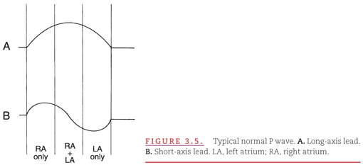
The P-wave contour is normally smooth and is either entirely positive or entirely negative (see Fig. 1.9; monophasic) in all leads except V1 and possibly V2. In the short-axis view provided by lead V1, which best distinguishes left- versus right-sided cardiac activity, the divergence of right- and left-atrial activation typically produces a biphasic P wave (see Fig. 1.14). The contributions of right- and left-atrial activation to the beginning, middle, and end of the P wave are indicated in Figure 3.5. Typical appearances of a normal P wave in a long-axis lead such as II (see Fig. 3.5A) and a short-axis lead such as V1 (see Fig. 3.5B) are illustrated.
P-Wave Duration
The P-wave duration is normally <0.12 second. Displayed in Figure 3.5, the P-wave duration is divided into thirds (vertical lines) to indicate the relative times of activation in the right and left atria.
Positive and Negative Amplitudes
The maximal P-wave amplitude is normally no more than 0.2 mV in the frontal plane leads and no more than 0.1 mV in the transverse plane leads.
Axis in the Frontal and Transverse Planes
The P wave normally appears entirely upright in leftward- and inferiorly oriented leads such as I, II, aVF, and V4 to V6. It is negative in aVR because of the rightward orientation of that lead and is variable in other standard leads. The direction of the P wave, or its axis in the frontal plane, should be determined according to the method for determining the axis of an ECG waveform presented later in the section “Morphology of the QRS Complex.” The normal limits of the P-wave axis are between 0 degrees and +75 degrees.1
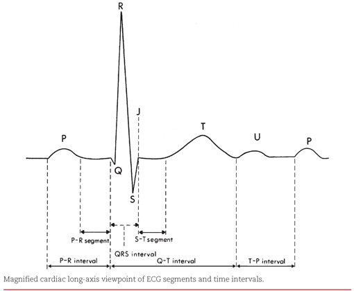
The PR interval measures the time required for an electrical impulse to travel from the atrial myocardium adjacent to the sinoatrial (SA) node to the ventricular myocardium adjacent to the fibers of the Purkinje network (see Fig. 1.12, repeated here). This duration is normally from 0.10 to 0.21 second. A major portion of the PR interval reflects the slow conduction of an impulse through the atrioventricular (AV) node, which is controlled by the balance between the sympathetic and parasympathetic divisions of the autonomic nervous system. Therefore, the PR interval varies with the heart rate, being shorter at faster rates when the sympathetic component predominates, and vice versa. The PR interval tends to increase with age2:
In childhood: 0.10 to 0.12 second
In adolescence: 0.12 to 0.16 second
In adulthood: 0.14 to 0.21 second
In developing a systematic approach to waveform analysis, the following steps should be taken to determine the morphology of the QRS complex.
General Contour
The QRS complex is composed of higher frequency signals than are the P and T waves, thereby causing its contour to be peaked rather than rounded. Positive and negative components of the P and T waves are simply termed positive and negative deflections, whereas those of the QRS complex are assigned specific labels, such as “Q wave” (see Fig. 1.10).
Q Waves
In some leads (V1, V2, and V3), the presence of any Q wave should be considered abnormal, whereas in all other leads (except rightward-oriented leads III and aVR), a “normal” Q wave is very small. The upper limit of normal for such Q waves in each lead is illustrated in Figure 3.6 and indicated in Table 3.1.3

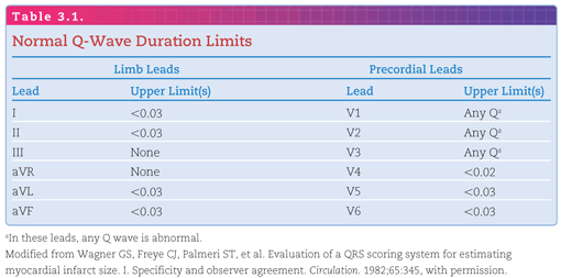
The absence of small Q waves in leads V5 and V6 should be considered abnormal. A Q wave of any size is normal in leads III and aVR because of their rightward orientations (see Fig. 2.4). Q waves may be enlarged by conditions such as local loss of myocardial tissue (infarction), enlargement (hypertrophy or dilatation) of the ventricular myocardium, or abnormalities of ventricular conduction.
R Waves
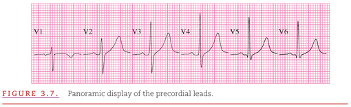
Because the precordial leads provide a panoramic view of the cardiac electrical activity progressing from the thinner right ventricle across the thicker left ventricle, the positive R wave normally increases in amplitude and duration from lead V1 to lead V4 or V5 (Fig. 3.7). Reversal of this sequence, with larger R waves in leads V1 and V2, can be produced by right-ventricular hypertrophy, and accentuation of this sequence, with larger R waves in leads V5 and V6, can be produced by left-ventricular hypertrophy. Loss of normal R-wave progression from lead V1 to lead V4 may indicate loss of left-ventricular myocardium, as occurs with myocardial infarction (see Chapter 12).
S Waves
The S wave also has a normal sequence of progression in the precordial leads. It should be large in V1, larger in V2, and then progressively smaller from V3 through V6 (see Fig. 3.7). As with the R wave, this sequence could be altered by hypertrophy of one of the ventricles or myocardial infarction.
QRS Complex Duration
The duration of the QRS complex is termed the QRS interval, and it normally ranges from 0.07 to 0.11 second (see Fig. 1.12). The duration of the complex tends to be slightly longer in males than in females.4 The QRS interval is measured from the beginning of the first-appearing Q or R wave to the end of the last-appearing R, S, R′, or S′ wave. Figure 3.8 illustrates the use of three simultaneously recorded limb leads (I, II, and III) to identify the true beginning and end of the QRS complex. An isoelectric period of approximately 0.02 second is apparent in lead II at the beginning of the QRS complex, and an isoelectric period of approximately 0.01 second is apparent in lead III at the end of the QRS complex. Note that only lead I reveals the true QRS duration (0.12 second).

Such multilead comparison is necessary; either the beginning or the end of the QRS complex may be isoelectric (neither positive nor negative) in a particular lead, causing an apparently shorter QRS duration. This isoelectric appearance occurs whenever the summation of ventricular electrical forces is perpendicular to the involved lead. The onset of the QRS complex is usually quite apparent in all leads, but its ending at the junction with the ST segment (termed the J point) is often indistinct, particularly in the precordial leads. The QRS interval has no lower limit that indicates abnormality. Prolongation of the QRS interval may be caused by left-ventricular hypertrophy, an abnormality in intraventricular impulse conduction, or a ventricular site of origin of the cardiac impulse.
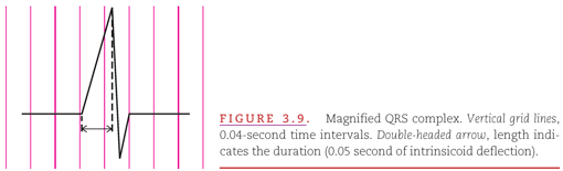
The duration from the beginning of the earliest appearing Q or R wave to the peak of the R wave in several of the precordial leads has been termed the intrinsicoid deflection (Fig. 3.9). Electrical activation of the myocardium begins at the endocardial insertions of the Purkinje network. The end of the intrinsicoid deflection represents the time at which the electrical impulse arrives at the epicardial surface as viewed by that particular lead. The deflection is called an intrinsic deflection when the electrode is on the epicardial surface and an intrinsicoid deflection when the electrode is on the body surface.5
Positive and Negative Amplitudes
Stay updated, free articles. Join our Telegram channel

Full access? Get Clinical Tree


