Fig. 8.2; Table 1 The ECG signs of right ventricular hypertrophy (RVH). The impact of RVH is on the QRS axis (which shifts to the right (a) and the right-sided chest leads (b)); there is no impact on the left-sided chest leads. As RVH advances, the size of the R wave in lead V1 gradually increases (ii), becoming dominant (iii). In advanced RVH, in addition to a ‘dominant’ R wave in lead V1, the T waves in the right-sided chest leads flattens and then inverts (‘strain’, or ‘repolarization changes’). Finally, right bundle branch block occurs (iv).
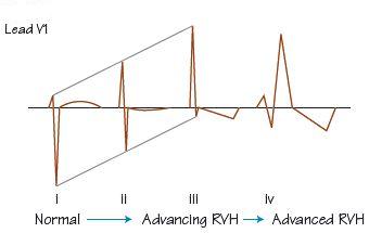
Fig. 8.3; Table 2 ECG consequences of advancing left ventricular hypertrophy (LVH). (a) As LVH progresses, increasing left axis deviation occurs, the height of the R wave in the left-sided leads increases (often with increasing size of the initial small Q wave in leads V5 and V6, due to increased voltages generated by left to right depolarization of the larger interventricular septum). The T wave in the lateral leads first flattens (ii), then inverts (see iv) (‘strain’ or ‘repolarization changes’). (b) In the right-sided leads, the S wave deepens, but there is no impact on the ST–T wave. In advanced LVH left bundle branch block often occurs (v). RVH, right ventricular hypertrophy.
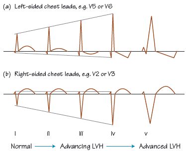
Table 1 Criteria for the ECG diagnosis of right ventricular hypertrophy (RVH).
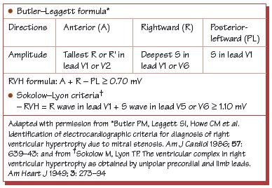
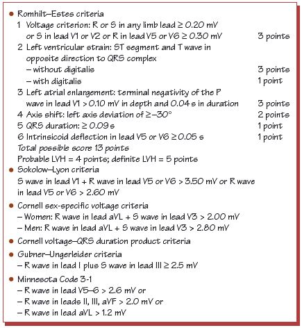
Table 2 Criteria for the ECG diagnosis of left ventricular hypertrophy (LVH).
The size of the QRS complex depends on:
- The number and activity of myocytes. Myocytes may become less numerous with age. Myocytes are more electrically active in youth (< 40 years) and in ventricular hypertrophy.
- The insulation between the heart and the observing electrodes. A pericardial effusion, or obesity, diminishes the amount of electricity reaching the electrodes. The latter is easily diagnosed, the former, either by clinical signs or, rarely, by beat-to-beat variation in the amplitude of the QRS complex (see Chapter 25).
Right ventricular hypertrophy
Right ventricular hypertrophy (RVH) results in greater voltages from the right ventricle (see Fig. 8.2), resulting in:
Stay updated, free articles. Join our Telegram channel

Full access? Get Clinical Tree


