Fig. 27.2 Anxiety-induced ST depression. Sinus tachycardia heart rate 150 b/min, with a P wave preceding each R wave. Normal P wave, PR interval, QRS complexes. Dramatic downward sloping ST depression laterally (V4–6), and inferiorly (II, III, aVF). There are many explanations of such an ECG. The patient was prone to anxiety, recovering from orthopaedic surgery when this ECG was taken; pulmonary emboli is a real possibility, as is myocardial ischaemia, and these must both be excluded. The diagnosis turned out to be anxiety induced ECG changes, with the immediate post-episode ECG being normal.
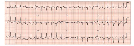
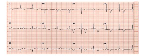
Fig. 27.3 Tako-tsubo syndrome. Woman with chest pain, small troponin rise. Pan-anterior (I, aVL, V1–6) deep symmetrical T wave inversion. High-grade left anterior descending (LAD) lesion must be angiographically excluded; the ventriculogram confirmed the typical contractile abnormality.
Emotion can affect the ECG
Emotion can affect the ECG two ways:
- Firstly, by causing asymptomatic ECG changes, leading to an erroneous diagnosis of heart disease. These changes are limited to ST/T wave changes (i.e. not involving the development of a Q wave, or changes to the QRS complex).
- Secondly, much more rarely, by causing ‘organic’ cardiac disease, such as ‘standard’ myocardial infarction, arrhythmias or the syndrome of transient left ventricular dysfunction.
Diagnostic difficulties
An extraordinary common difficult problem in clinical practice is to decide whether an abnormal ECG reflects cardiac pathology or not, with the ECG changes being explained by anxiety or hyperventilation.
Anxiety-related ECG changes
Anxiety can profoundly alter the ECG, probably via changes in autonomic nervous system function, as evidenced by the ECG normalizing with manoeuvres that normalize autonomic function (reassurance, rest, and anxiolytics and beta-blockers), with catecholamine infusion producing similar ECG changes. The ECG changes in anxiety are:
- ST flattening, the commonest finding.
- Frank ST depression; not rare, especially in hyperventilation.
- T wave inversion
How does one differentiate anxiety-induced changes from those reflecting cardiac disease? Fully assessing demographics (age, sex, etc.) and symptomatology are the keys to a correct diagnosis. Though one can often be fairly confident that some patterns are due to anxiety, one cannot always be 100% certain, so it is not uncommon to undertake further limited investigations to exclude heart disease, always explaining beforehand that the result is likely to negative, and the test is required only for reassurance.
ST elevation and emotion
Stay updated, free articles. Join our Telegram channel

Full access? Get Clinical Tree


