(1)
The Lindner Center for Research and Education The Christ Hospital, The Christ Hospital Physicians Ohio Heart and Vascular Center, Cincinnati, OH, USA
Keywords
Diagnostic performanceACCURACY trialCORE 64 trialPrognostic valueCONFIRM studyPlaque characteristicsZero calcium scorePromise StudySCOT Heart StudyCoronary computed tomographic angiography (CCTA) has become a high diagnostic performing noninvasive imaging modality when compared to invasive coronary angiography (ICA). Its particular strength is in its very high negative predictive value for the evaluation of obstructive coronary artery disease (CAD). In other words, its best use is to rule out CAD in low to intermediate risk patient populations. CCTA may serve as a gate keeper to ICA to curb unnecessary use. Studies have shown that up to 39 % of patients undergoing invasive coronary angiography have no significant CAD defined as >20 % luminal stenosis [1]. The following chapter is meant to highlight significant studies in the field that demonstrate the diagnostic performance and clinical utility of CCTA. The chapter is by no means meant to be inclusive of all CCTA studies.
Diagnostic Performance
The diagnostic performance of CCTA is comparable to other noninvasive coronary imaging modalities. A meta-analysis of 27 studies using 16–64-slice scanners reported a sensitivity, specificity, negative predictive value (NPV) and positive predictive value (PPV) of 99, 89, 93 and 100 % respectively [2]. Specifically, the ACCURACY trial (Assessment by Coronary Computed Tomographic Angiography of Individuals Undergoing Invasive Coronary Angiography) was the first multicenter study. It evaluated 230 patients without known CAD undergoing CCTA before ICA [3]. ACCURACY demonstrated a sensitivity, specificity, PPV and NPV of 95, 83, 64 and 99 % respectively. Two additional individual trials including a 27 study meta-analysis performed subsequent to ACCURACY reported similar results. These were the CORE64 (CORonary artery Evaluation using 64-row multidetector CT angiography) Trial [4] and the European trial [5]. One other real world study was published with similar findings [6]. See Fig. 8.1.
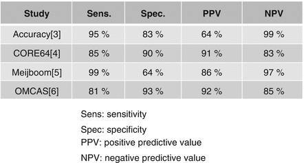
Figure 8.1
The diagnostic accuracy of coronary computed tomography angiography
An additional advantage of CCTA is its ability to characterize plaque independent of its luminal stenosis grade. A meta-analysis study of 33 trials comprising 946 patients demonstrated a good correlation between intravascular ultrasound (IVUS) and CCTA for atherosclerosis measures above and beyond stenosis grade [7]. These factors include plaque cross sectional area, plaque area, area stenosis, plaque volume and arterial remodeling [7].
Prognostic Value of CCTA
Mortality
In addition to diagnostic accuracy, CCTA has been determined to add incremental clinical prognostic value. The first large study evaluating the prognostic value of CCTA was a single center trial evaluating the association of all-cause mortality with CCTA defined extent and degree of CAD in 1,127 patients with suspected CAD [8]. Patients were followed for 15 months. In this study, 16-row detector CCTA data was scored for stenosis severity, plaque burden and clinical grading scores for plaque extent and distribution. CCTA independent predictors of all death included proximal left anterior descending artery stenosis and number of vessels with ≥50 % and ≥70 % stenoses. Survival worsened with greater number of vessels with ≥70 % stenosis. Survival with 1 stenosis ≥70 % or 2 stenoses of 50 % was 96 %. Whereas, survival for left main stenosis of ≥50 % was 86 %. Negative CCTA findings proved to portend very low clinical risk of death. Since this study, multiple other studies have evaluated the prognostic significance of CCTA. Min et al. in a large scale 2 center study examined 5,330 consecutive patients without known CAD who underwent CCTA [9]. These patients were followed for all cause mortality for a little over 2 years. Obstructive CAD of > 70 % correlated with a high incidence of death with a hazard ratio (HR) of 2.44, 95 % confidence interval: 1.61–3.72 (p < 0.0001). Compared with those without obstructive CAD, individuals with increasing numbers of vessels with obstructive CAD suffered from a greater incidence of death (1 vessel, HR 2.23, 95 % confidence interval: 1.34–3.73; 2 vessels, HR 3.29, 95 % confidence interval: 1.62–6.71; 3 vessel, HR 7.35, 95 % confidence interval: 3.79–14.29; p, 0.001 for all). Additionally, ejection fraction performed using retrospective imaging by CCTA also proved to have prognostic value. Left ventricular ejection fraction (LVEF) <50 % versus those >50 % exhibited higher mortality rates (HR 1.56, 95 % confidence interval: 1.04–2.36, p = 0.03). Finally, annualized mortality rates for those with obstructive CAD and depressed LVEF were higher than those with obstructive CAD and normal LVEF. Those with nonobstructive or no CAD and normal LVEF had the lowest mortality rates. Those with nonobstructive CAD and normal LVEF faired better than those with nonobstructive CAD and low LVEF.
In addition, the CONFIRM [10] study demonstrated that in risk-adjusted analyses, both obstructive and nonobstructive CAD predicted an increased risk of mortality compared to those without CAD (HR 2.60, 95 % confidence interval: 1.94–3.49, p, 0.0001 and HR 1.60, 95 % confidence interval: 1.18–2.16, p = 0.002 respectively). Additionally, it was noted that all-cause mortality increased with increasing number of coronary vessels involved. For nonobstructive CAD, obstructive 1 vessel disease, obstructive 2 vessel disease and obstructive 3 vessel or left main disease the hazard ratios were (1.62, 95 % confidence interval: 1.20–2.19, p = 002; 2.00, 95 % confidence interval: 1.43–2.82, p < 0.0001; 2.92, 95 % confidence interval: 2.00–4.25, p < 0.0001; 3.70, 95 % confidence interval: 2.58–5.29, p < 0.0001 respectively). The absence of coronary artery disease demonstrated a low annual mortality rate of 0.28 %. The hazard ratio for death among females with obstructive CAD was higher than that for males with obstructive CAD (4.2, 95 % confidence interval: 2.47–7.18, p < 0.0001 versus 3.27, 95 % confidence interval: 1.96–5.45, p < 0.0001).
The CONFIRM registry has also demonstrated that individuals with diabetes mellitus have a higher frequency of obstructive CAD compared to nondiabetic patients as evaluated by CCTA [11]. Additionally, the risk of mortality was significantly higher in diabetics than those without diabetes for those with no CAD, those with nonobstructive CAD and those with obstructive 1 vessel CAD, obstructive 2 vessel CAD and obstructive 3 vessel CAD [11].
Finally reduced LVEF (EF < 50 %), measured by CCTA, has been reported to portend an increased mortality versus those with normal EF [12].
Major Adverse Cardiovascular Events
Regarding the prognostic value of CCTA for major adverse cardiovascular outcomes (MACE), there have been only a few studies, most of which were small, single center trials. One larger study by Hadamitzky et al. [13] prospectively evaluated the prognostic utility of CCTA in 1,256 patients with suspected CAD for the occurrence of MACE (cardiac death, myocardial infarction, unstable angina and target vessel revascularization > 90 days after CCTA). In the 802 patients without obstructive CAD, there were 4 MACE events versus 17 MACE events in the 348 patients with obstructive CAD, which was highly significant (p < 0.001). Another meta-analysis of 10 studies using 64-slice CCTA showed a cumulative MACE rate of 0.5 % in patients with normal CCTA, 3.5 % in those with nonobstructive CAD and 16 % with obstructive CAD [14].
< div class='tao-gold-member'>
Only gold members can continue reading. Log In or Register to continue
Stay updated, free articles. Join our Telegram channel

Full access? Get Clinical Tree


