Definitions of Acute Coronary Syndromes: Introduction
Coronary heart disease (CHD) is a worldwide health epidemic. In the United States, for example, it is estimated that 13.7 million Americans have CHD, including more than 7.2 million individuals who already have had a myocardial infarction (MI).1 In the group of persons older than 30 years of age, 213 per 100,000 individuals have CHD.1 Although age-specific events related to CHD have fallen dramatically in the last few decades, the overall prevalence has risen as populations age and patients survive the initial coronary or cardiovascular event. The Centers for Disease Control and Prevention estimates that life expectancy in America might be increased by 7 years if CHD and its complications were eradicated.2 Worldwide, 30% of all deaths can be attributed to cardiovascular disease, of which more than half are caused by CHD, and the forecasts for the future estimate a growing number as a consequence of lifestyle changes in developing countries.2 Globally, of those dying from cardiovascular diseases, 80% are in developing countries and not in the Western world.2
CHD represents a continuum of disease pathologies and its subsequent risks. CHD has been classified as chronic CHD, acute coronary syndromes, and sudden death. CHD may present clinically in many ways, extending from an asymptomatic finding to unexpected cardiac collapse. Chronic CHD is always secondary to coronary atherosclerosis, leading to mismatch of coronary blood flow and adenosine triphosphate homeostasis (imbalance of supply and demand) and a stable pattern of coronary ischemia. The clinical pattern includes stable angina pectoris and myocardial hibernation.3 This chapter, however, focuses on a more high-risk population, those with acute coronary syndromes.
Acute Coronary Syndromes
Acute coronary syndrome (ACS) is a unifying term representing a common end result, acute myocardial ischemia. Acute ischemia is usually, but not always, caused by atherosclerotic plaque rupture, fissuring, erosion, or a combination with superimposed intracoronary thrombosis and is associated with an increased risk of cardiac death and myonecrosis.4 It encompasses acute MI (resulting in ST-segment elevation or non–ST-segment elevation) and unstable angina. Recognizing a patient with ACS is important because the diagnosis triggers both triage and management. Those deemed to have an acute coronary syndrome in the emergency department should be triaged immediately to an area with continuous electrocardiographic monitoring and defibrillation capability. An electrocardiogram (ECG) should be obtained and accurately interpreted within 10 minutes. Those patients with suspected ACS should be managed immediately with antiplatelet and anticoagulant therapies and considered for immediate revascularization mechanically or pharmacologically if new ST-segment elevation is noted.5
Because of the life-threatening nature of an ACS, it is prudent to have a low threshold in suspecting a patient with acute chest pain as potentially having an ACS. Because the efficient diagnosis and optimal management of these patients are derived from information mostly only readily available from initial clinical presentation, there is overlap of those with true ACS and those who ultimately do not have CHD as a cause of their cardiac symptoms. In addition, it may not be possible to differentiate patients with MI (either ST-segment elevation or non–ST-segment elevation) from those with unstable angina in the initial hours, as the biomarkers of myonecrosis can be normal initially.
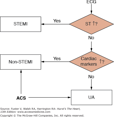
Nonetheless, proper initial triage of patients suspected to have acute coronary ischemia should eventually identify patients as having (1) ACS; (2) a non-ACS cardiovascular condition such as myocarditis/myopericarditis, stress-related cardiomyopathy, aortic dissection, or pulmonary embolism; (3) a noncardiac cause of chest pain such as gastroesophageal reflux; and (4) a noncardiac condition that is yet undefined, such as sepsis.6 ACS patients with new evidence of ST-segment elevation on the presenting ECG are labeled as having an ST-segment elevation MI (STEMI) and should be considered for immediate reperfusion therapy by thrombolytics or percutaneous coronary intervention (PCI); those without ST-segment elevation but with evidence of myonecrosis are deemed to have a non–ST-segment elevation MI (NSTEMI); and those without any evidence of myonecrosis are diagnosed with unstable angina (Fig. 56–1).
Definition of Unstable Angina
Unstable angina is usually secondary to reduced myocardial perfusion resulting from coronary artery atherothrombosis. In this event, however, the nonocclusive thrombus that developed on a disrupted atherosclerotic plaque does not result in any biochemical evidence of myocardial necrosis. Unstable angina and NSTEMI can be viewed as very closely related clinical conditions with similar presentations and pathogenesis but of differing severity.
Because of the lack of objective data associated with the condition, unstable angina (also known as preinfarction angina, intermediate coronary syndrome, and acute coronary insufficiency) must be diagnosed from careful history taking and is thus the most subjective of the ACS diagnoses. The Agency for Health Care Policy and Research has published guidelines listing features that signify the likelihood of signs and symptoms suggestive of an ACS likely caused by CHD (Table 56–1).7 There are three principal presentations of unstable angina: (1) rest angina or angina with minimal exertion usually lasting at least 20 minutes; (2) new-onset severe angina, usually defined as occurring within the last month; and (3) crescendo angina, defined as previously diagnosed angina that has become distinctly more frequent, longer in duration, or more severe in nature.8
| Feature | High Likelihood: Any of the Following | Intermediate Likelihood: Absence of High Likelihood Features and Presence of Any of the Following | Low Likelihood: Absence of High- or Intermediate-Likelihood Features But May Have the Following |
|---|---|---|---|
| History | Chest or left arm pain or discomfort as chief symptom reproducing documented angina; known history of CAD, including MI | Chest or left arm pain or discomfort as chief symptom; age >70 years; male sex; diabetes mellitus | Probable ischemic symptoms in absence of any of the intermediate likelihood characteristics; recent cocaine use |
| Examination | Transient MR, hypotension, diaphoresis, pulmonary edema, or rales | Extracardiac vascular disease | Chest discomfort reproduced by palpation |
| ECG | New, or presumably new, transient, ST-segment deviation (≥ 0.05 mV), or T-wave inversion (≥ 0.2 mV) with symptoms | Fixed Q waves; abnormal ST segments or T waves not documented to be new | T-wave flattening or inversion in leads with dominant R waves; normal ECG |
| Cardiac markers | Elevated cardiac TnI, TnT, or CK- MB | Normal | Normal |
Because of the heterogenous nature of the patients who fall under these loose definitions, many classification schemes have been proposed for unstable angina. Although not devised precisely to help define unstable angina, the Canadian Cardiovascular Society (CCS) has developed an easy classification system to grade anginal symptoms (Table 56–2).9 Class I angina is the least symptomatic and denotes that ordinary physical activity does not illicit anginal symptoms. Class II angina implies anginal symptoms that slightly impair ordinary activity such as walking >2 blocks or climbing >1 flight of stairs. Class III angina is defined as symptoms that limit markedly ordinary physical activity such as walking <1 block or climbing <1 flight of stairs. Finally, Class IV angina is symptoms at rest or that cause an inability to carry on any physical activity without discomfort. Using this classification, crescendo angina can be defined as symptoms that result in at least 1 CCS class increase or to at least CCS class III severity.10
| Class | Description of Stage |
|---|---|
| I | “Ordinary physical activity does not cause…angina,” such as walking or climbing stairs. Angina occurs with strenuous, rapid, or prolonged exertion at work or recreation. |
| II | “Slight limitation of ordinary activity.” Angina occurs on walking or climbing stairs rapidly; walking uphill; walking or stair climbing after meals; in cold, in wind, or under emotional stress; or only during the few hours after awakening. Angina occurs on walking >2 level blocks and climbing >1 flight of ordinary stairs at normal pace and under normal conditions. |
| III | “Marked limitation of ordinary physical activity.” Angina occurs on walking 1 to 2 level blocks and climbing 1 flight of ordinary stairs under normal conditions and at normal pace. |
| IV | “Inability to carry on any physical activity” without discomfort—anginal symptoms may be present at rest. |
Braunwald developed a useful classification of unstable angina by assessing risk.11 By differentiating the severity and clinical circumstances surrounding the presentation of unstable angina and considering also the presence or absence of ECG changes and the intensity of medical therapy, Braunwald has estimated the risk of death or MI at 1 year. In terms of severity, class I unstable angina is new onset or accelerated angina but with no rest pain. Class II presents with rest angina within the last month but not within the previous 48 hours. Class III angina presents at rest and within the last 48 hours of initial evaluation. In terms of clinical circumstances, class A represents unstable angina in the setting of a secondary noncoronary cause of demand ischemia such as anemia, hypotension, or prolonged tachycardia. Class B is worsening primary CHD in the absence of extracardiac conditions. Class C is postinfarction unstable angina within 2 weeks of a documented MI (Table 56–3). Furthermore, patients fared worse over the following 12 months if they presented with transient ST-T–wave changes during pain and if they had angina despite maximal anti-ischemic therapy. In summary, patients with a 48-hour pain-free interval and the absence of ECG changes were at decreased risk, whereas those with postinfarction angina and the need for maximal medical therapy have the highest risk of death or MI over the next 1 year after presentation with unstable angina.
| Clinical Circumstances | |||
|---|---|---|---|
| Severity | A. Develops in Presence of Extracardiac Condition that Intensifies Myocardial Ischemia (Secondary UA) | B. Develops in Absence of Extracardiac Condition (Primary UA) | C. Develops Within 2 Weeks After Acute MI (Postinfarct UA) |
| I | IA | IB | IC |
| II | IIA | IIB | IIC |
| III | IIIA | IIIB | IIIC |
The Agency for Health Care Policy and Research also has published guidelines assessing the short-term risk of death or nonfatal MI in patients with unstable angina using similar clinical features (Table 56–4). It should be noted that an elevated level of a cardiac marker such as a troponin places the patient at high risk. These patients would now be considered to have an NSTEMI instead of high-risk unstable angina. The risk of death at 6 months with unstable angina is approximately 3% versus 9% with NSTEMI, illustrating the importance of an accurate diagnosis.11
| Feature | High Risk: At Least 1 of the Following Features Must be Present | Intermediate Risk: No High-Risk Feature But Must Have 1 of the Following | Low Risk: No High- or Intermediate-Risk Feature But May Have Any of the Following |
|---|---|---|---|
| History | Accelerating tempo of ischemic symptoms in preceding 48 h | Prior MI, peripheral or cerebrovascular disease or CABG, prior aspirin use | |
| Character of pain | Prolonged ongoing (>20 min) rest pain | Prolonged (>20 min) rest angina, now resolved, with moderate or high likelihood of CAD; rest angina (<20 min) or relieved with rest or sublingual NTG | New-onset or progressive CCS class III or IV angina within the past 2 wk without prolonged (>20 min) rest pain but with moderate or high likelihood of CAD (see Table 56–1). |
| Clinical findings | Pulmonary edema, most likely caused by ischemia; new or worsening MR murmur; S3 or new/worsening rales; hypotension, bradycardia, tachycardia; age >75 y | Age >70 y | |
| ECG | Angina at rest with transient ST-segment changes >0.05 mV; bundle-branch block, new or presumed new; sustained ventricular tachycardia | T-wave inversions >0.2 mV; pathologic Q waves | Normal or unchanged ECG during an episode of chest discomfort |
| Cardiac markers | Elevated (eg, TnT or TnI >0.1 ng/mL) | Slightly elevated (eg, TnT >0.01 but <0.1 ng/mL) | Normal |
Although nonocclusive thrombus on a preexisting atherosclerotic plaque is the most common cause of unstable angina/NSTEMI, other causes may lead to acute coronary ischemia (Table 56–5).6 A less common cause is dynamic obstruction of an epicardial artery leading to intense focal spasm (Prinzmetal angina). It is thought that this spasm is caused by hypercontractility of vascular smooth muscle and/or endothelial dysfunction. Abnormal constriction of small intramural resistance vessels can also lead to dynamic obstruction and acute ischemia. A third cause of unstable angina is severe mechanical obstruction without spasm or thrombus. An example would be restenosis after PCI or some patients with progressive atherosclerosis. A fourth cause is arterial inflammation and/or infection. It is thought that chronic inflammation perhaps related to infection lead to activation of macrophages and T-lymphocytes at the shoulder of a vulnerable plaque and increased expression of metalloproteinase resulting in disruption and rupture of the plaque. Finally, a fifth cause of unstable angina is alluded to in Braunwald’s classification, namely unstable angina from a secondary cause. These patients generally have chronic, stable CHD that worsens as a result of a noncoronary condition that increases myocardial oxygen demand, such as fever, tachycardia, reduced coronary blood flow caused by hypotension, and reduced blood myocardial oxygen content, such as with hypoxemia or anemia. These causes are not mutually exclusive.
| Increased myocardial oxygen demand |
Fever Thyrotoxicosis Tachycardia Malignant hypertension Pheochromocytoma Aortic stenosis High output state Pregnancy Drugs, cocaine, amphetamine |
| Decreased oxygen supply |
Anemia Hypoxemia Carbon monoxide poisoning Polycythemia vera Hyperviscosity syndromes |
Non–ST-Segment Elevation Myocardial Infarction
For many years, the diagnosis of an acute MI was defined by the World Health Organization as having two of the following three criteria: (1) typical ischemic chest pain; (2) typical ECG pattern including the development of Q waves; (3) typical rise and fall in serum markers of myocardial injury, usually creatine kinase myocardial band (CK-MB).12 If the patient did not have evidence of ST-segment elevation or Q waves, and the CK-MB was elevated, the patient was diagnosed with an NSTEMI. Those patients with acute ischemic chest pain without evidence of ST-segment elevation or Q waves and who had negative CK-MB levels were thought to have unstable angina.
With the introduction of serum troponin levels, which were much more sensitive and specific for myonecrosis than CK-MB levels, a joint European Society of Cardiology and American College of Cardiology committee convened and in 2000 proposed the following definition of an acute, evolving, or recent MI: Typical rise and gradual fall of serum troponin levels or a more rapid rise and fall of serum CK-MB levels in addition to presenting with either ischemic symptoms, development of pathologic Q waves on ECG, ST-segment changes indicative of ischemia, or coronary artery intervention (eg, PCI).13
Patients presenting with NSTEMI have an intermediate risk of acute complications when compared with patients with unstable angina (lower risk) and STEMI (higher risk), with a 30-day mortality rate of approximately 5%.11
Stay updated, free articles. Join our Telegram channel

Full access? Get Clinical Tree


