Coronary Stenting


Coronary Stenting


Ajay J. Kirtane

Gregg W. Stone


Stents are metallic scaffolds that are deployed within diseased segments of coronary arteries to establish and maintain wide luminal patency. Currently, stent-assisted coronary intervention is the most common revascularization modality in patients with coronary artery disease. The acute and late results of stent implantation, however, vary greatly depending on the clinical risk profile of the patient, the complexity of the coronary lesion and interventional procedure, and the specific stent device that is used. A broad range of evidence is available from clinical trials conducted over the past two decades to guide appropriate stent usage in most situations. The present chapter traces the evolution and development of the coronary stent from its initial applications to treat balloon angioplasty failures to its widespread global adoption for the treatment of patients with ischemic coronary heart disease.


BARE-METAL STENT OVERVIEW


Limitations of Balloon Angioplasty

While the performance of the first successful balloon angioplasty on September 16, 1977, in Zurich, Switzerland, set the stage for the millions of percutaneous coronary intervention (PCI) procedures that have since taken place, stand-alone balloon angioplasty as performed by Andreas Gruentzig and other early pioneers was a highly unpredictable experience. The mechanism of balloon angioplasty involves plaque fracture (dissection) into the deep media, with expansion of the external elastic lamina, as well as partial axial plaque redistribution along the length of the treated vessel. The majority of vessels undergoing balloon angioplasty tolerate balloon dilatation and heal sufficiently to result in an adequate lumen; however, balloon-mediated injury to the vessel wall can at times be uncontrolled and excessive, resulting in balloon angioplasty’s two major limitations: abrupt closure (occurring acutely, or within the first several days after angioplasty) and restenosis (occurring later, within months after the procedure due to a combination of acute recoil and chronic constrictive remodeling). The coronary stent was thus devised as an endoluminal scaffold to create a larger initial lumen, to seal dissections, and to resist recoil and late vascular remodeling, thereby improving upon the early and late results of balloon angioplasty.


Development of the Coronary Stent

The term “stent” derives from a dental prosthesis developed by the London dentist Charles Stent (1807-1885) and is now used to indicate any device used for “extending, stretching, or fixing in an expanded state”.1 The first stents were implanted in human coronary arteries in 1986 by Ulrich Sigwart, Jacques Puel, and colleagues, who placed the Wallstent sheathed self-expanding metallic mesh scaffold (Medinvent, Lausanne, Switzerland) in the peripheral and coronary arteries of eight patients.2 Further experience with this device demonstrated high rates of thrombotic occlusion and late mortality,3 although patients without thrombosis had a 6-month angiographic restenosis rate of only 14%, suggesting for the first time that stenting could improve late patency in addition to stabilizing the acute results obtained after conventional balloon angioplasty. Another early stent platform developed contemporaneously by Cesare Gianturco and Gary Roubin was a balloon-expandable coil stent consisting of a wrapped stainless steel wire resembling a clamshell (Figure 31.1, left). A phase II study evaluating the Gianturco-Roubin stent to reverse postangioplasty acute or threatened vessel closure was started in 1988,4 ultimately leading to United States Food and Drug Administration (FDA) approval for this indication in June 1993.

While these stents were being developed and tested, Julio Palmaz designed a balloon-expandable slotted tube stainless steel stent in which rectangular slots were cut into thinwalled stainless steel tubing and deformed into diamondshaped windows during expansion by an underlying delivery
balloon. While this design allowed for relatively straightforward deployment, the rigidity of this initial stent design made it difficult to deliver this device to the coronary vasculature. In 1989 a design modification was made by Richard Schatz, consisting of the placement of a 1 mm central articulating bridge connecting the two rigid 7 mm slotted segments,5 creating the 15 mm Palmaz-Schatz stent (Johnson and Johnson Interventional Systems, Warren, NJ) (Figure 31.1, right). The first coronary Palmaz-Schatz stent was placed in a patient by Eduardo Sousa in São Paulo, Brazil in 1987 with a US pilot study started in 1988.


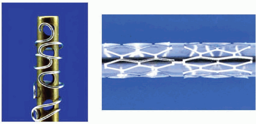
Figure 31.1 Left. The Gianturco-Roubin Stent. Stainless steel sutures were wound around a cylindrical rod using pegs to shape the wire, resulting in a clamshell design. Right. The Palmaz-Schatz Stent. Note the articulation between the two slotted tubes.

In 1989, enrollment commenced in two randomized multicenter studies (STRESS and BENESTENT) comparing balloon angioplasty alone to elective Palmaz-Schatz stenting. In these studies, the use of the Palmaz-Schatz stent was associated with a 20% to 30% reduction in clinical and angiographic restenosis compared with conventional balloon angioplasty (Figure 31.2).6,7 The Palmaz-Schatz stent also resulted in markedly improved initial angiographic results, with a larger postprocedural minimal luminal diameter and fewer residual dissections, which translated into a lower rate of subacute vessel closure. These results led to approval
of the Palmaz-Schatz stent by the FDA in 1994. Long-term follow-up up to 15 years has subsequently demonstrated few late clinical or angiographic recurrences from years 1 to 5 after coronary stent implantation,8,9 with slight and progressive decrements in luminal size thereafter extending beyond 10 years.10 The mechanisms of this late progression of disease are not entirely known, but have been hypothesized to be related to the development of new atherosclerosis within the originally stented segment rather than clot formation, as overall stent thrombosis rates have remained low (1.5% at 15 years).10


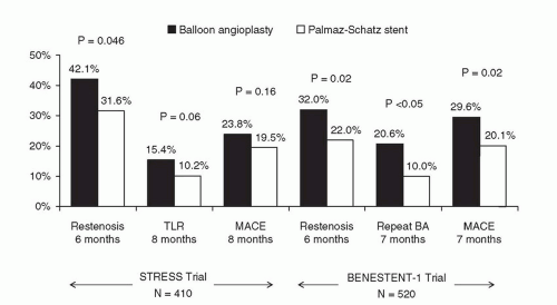
Figure 31.2 Results of STRESS and BENESTENT-1 landmark trials of the Palmaz-Schatz stent, which provided the evidence base for FDA approval of the Palmaz-Schatz stent for the prevention of restenosis in de novo lesions. BA, balloon angioplasty; TLR, target lesion revascularization; MACE, major adverse cardiac events.

Despite the impressive acute and long-term results with the Palmaz-Schatz stent which became the dominant stent design for coronary use, widespread adoption of stent technology was initially hindered by the perceived need for an intense anticoagulation regimen (consisting of aspirin, dipyridamole, heparin, dextran, and warfarin) to inhibit stent thrombosis (which nonetheless occurred in approximately 3% of patients). This profound degree of anticoagulation, however, resulted in a marked increase in hemorrhagic and vascular complications. It was not until further refinements in stent deployment technique and the utilization of dual antiplatelet therapy demonstrated reductions in these complications that stent usage became more widespread. Pioneers such as Antonio Colombo demonstrated reduced rates of stent thrombosis with more aggressive intravascular ultrasound (IVUS)-guided deployment techniques including routine high-pressure adjunctive dilatation (>14 atmospheres),11 along with the use of aspirin and a second antiplatelet agent (thienopyridine, ticlopidine) rather than prolonged warfarin therapy. These modifications significantly reduced the incidence of stent thrombosis to ˜1% to 2%, and concomitantly reduced bleeding and femoral arterial complications.12 The confirmation of Colombo’s initial findings in several randomized clinical trials (Figure 31.3) definitively established the superiority of dual antiplatelet therapy (with aspirin and ticlopidine) over an anticoagulation-based approach for prevention of stent thrombosis, and facilitated widespread adoption for coronary stenting by the late 1990s.13, 14, 15, 16


Stent Design: Impact on Performance and Clinical Outcomes


Classification

Coronary stents may be classified based on their composition (e.g., metallic or polymeric), configuration (e.g., slotted tube versus coiled wire), bioabsorption (either inert (biostable or durable) or degradable [bioabsorbable]), coatings (either none, passive such as heparin or polytetrafluoroethylene, PTFE), or bioactive (such as those eluting rapamycin or paclitaxel), and mode of implantation (e.g., self-expanding or balloon-expandable). The ideal stent would be made of a nonthrombogenic material and have sufficient flexibility in its unexpanded state to permit ready passage through guiding catheters and tortuous vessels, and yet have an expanded configuration providing uniform scaffolding of the vessel wall with low recoil and maximal radial strength while conforming to vessel bends. In addition, the ideal stent would be sufficiently radiopaque to allow fluoroscopic visualization to guide accurate placement and management of in-stent restenosis, but not so opaque as to obscure important angiographic vessel
details. In recent years, the importance of the stent delivery system to device profile, flexibility, and trackability around tortuous and calcific coronary vessels has received increasing appreciation. For balloon-expandable stents, the stent must be tightly crimped to the delivery balloon to avoid dislodgment, and the overhang of the balloon beyond the ends of the stent should be minimized (<1 mm) to avoid vessel trauma outside the stent margins. Stent delivery balloons must be able to withstand high pressures (>18 atm) without rupture, and should take into account a balance between deliverability versus a desire for low compliance to facilitate predictable sizing and avoid excessive growth outside the stent edges.


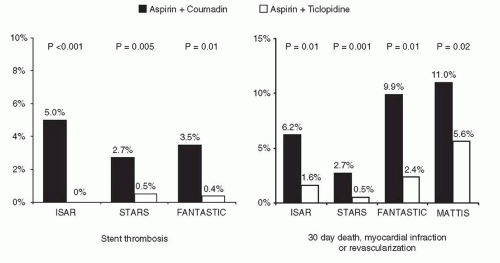
Figure 31.3 Benefits of dual antiplatelet therapy in reducing clinical events post stenting. Shown are the results from four landmark trials demonstrating the efficacy of antiplatelet (over antithrombotic) therapy.


Stent Composition

Until recently, the most widely used stent material was 316L stainless steel. Cobalt chromium and platinum chromium alloys have been employed in more recent stent designs in order to allow lower-profile thin stent struts (˜75 µm, versus 100 to 150 µm in most stainless steel stents) that still maintain radial strength and visibility. Most self-expanding stents utilize nitinol, a nickel/titanium alloy that has superelastic and thermal shape memory properties that allow it to be set into a particular expanded shape by baking at high temperature. Nitinol stents can then be squeezed down and constrained on the delivery system, able to return to that set shape when released in the coronary artery.

Other than gold (which has been shown to increase restenosis), there is little evidence that thrombosis or restenosis rates vary with the specific stent metal, though the final stages of surface finishing, smoothing, and purification or passivation may affect early thrombotic and late restenotic processes.17 There is a burgeoning interest in biodegradable stents, which theoretically offer the advantages of increased longitudinal flexibility (though at the expense of radial force), compatibility with noninvasive imaging, and complete bioabsorption over a period of months to a year or longer, thereby restoring underlying vascular reactivity. Bioabsorbable stents (or bioabsorbable scaffolds) are typically either polymeric in nature (e.g., using proprietary biodegradable polymers or poly-L-lactic acid (PLLA), which is degraded via the Krebs cycle to carbon dioxide and water) or nonpolymeric (e.g., magnesium-based).


Stent Configuration and Design

Stents can be assigned to one of three subgroups, based on construction: wire coils, slotted tubes/multicellular, and modular designs. After early experiences with wire coil stents (e.g., the Gianturco-Roubin stent), these types of stent designs rapidly fell out of favor because they in general lacked axial and radial strength, and due to lesser strut coverage predisposed to plaque prolapse. Thus, the vast majority of stents in current use are either slotted tube/multicellular or modular in design. In an effort to preserve the radial strength and wall coverage of the original tubular designs (e.g. the Palmaz stent) but improve flexibility in their collapsed states, several generations of slotted tube and multicellular stents have been introduced by various manufacturers. Each is laser cut from a metallic tube into a unique pattern that increases the overall flexibility of the stent by distributing bending throughout the stent length without compromising radial strength or elastic recoil in the expanded state. The newer stents are manufactured in a broad range of stent lengths (8 to 48 mm) and diameters (2.25 to 6.0 mm and above for peripheral applications) to facilitate stenting of long lesions, small vessels, saphenous vein grafts (SVGs), and distal lesions. To eliminate the need for a protective sheath, various mechanical, balloon-wrapping, and heat-curing processes have been developed to tightly crimp the stent onto the balloon until it is deployed. This bare mounting onto the delivery balloon has greatly reduced stent delivery profiles, comparable with the best angioplasty balloons of the late 1990s, and has kept stent embolization rates below approximately 1 to 3 per 1,000 procedures.

Despite their enhanced flexibility, even the latest-generation slotted tube stents are sometimes difficult to deliver through tortuous and noncompliant vessels. In an effort to enhance flexibility and deliverability without sacrificing the excellent scaffolding of the slotted tube stents, modular or hybrid stents have been created by flexibly joining multiple short repeating modules to each other. The initial modular stent was the Arterial Vascular Engineering MicroStent (subsequently purchased by Medtronic Corp., Santa Rosa, CA), which had a series of 4-mm-long, rounded stainless steel corrugated ring subunits welded to each other. Subsequent designs have incorporated an elliptorectangular (rounded) strut profile and progressively reduced the length of the individual modules, with progressive reductions in crossing profile and increased surface area coverage. Additionally, variation in the location and frequency of the weld-points has been used to engineer flexibility without attempting to sacrifice radial and axial strength.

Depending on the cellular configuration, multicellular stents can be broadly subclassified as either open cell or closed cell. Open cell designs tend to have varying cell sizes and shapes along the stent, and provide increased flexibility, deliverability, and side branch access by staggering the crosslinking elements to provide radial strength. Open cell designs thus tend to conform better on bends, though the cell area may open excessively on the outer curve of an angulated segment. Closed cell designs typically incorporate a repeating unicellular element that provides more uniform wall coverage with less tendency for plaque prolapse, at the expense of reduced flexibility and side branch access. Closed cell designs also tend to straighten vessel bends more than open cell designs.

Stent design may significantly impact acute and late vascular responses. Stents that possess better conformability, less rigidity, and greater circularity experimentally produce less vascular injury, thrombosis, and neointimal hyperplasia.18,19 Ex-vivo and clinical studies have suggested that thin stent
struts may be associated with reduced neointimal hyperplasia and lower rates of restenosis,20 in addition to inherently less thrombogenicity.21

Due to the recent emphasis upon thin-strutted and more flexible stent designs in order to facilitate deliverability as well as other adverse vascular responses to stent implantation, there have been some concerns regarding the integrity of modern stent platforms. While thin-strutted stents have obvious advantages, some of these stent platforms have been associated with a greater tendency for recoil (radial) or orthogonally, for axial (or “longitudinal”) deformation and/or compression.22,23 In the instance of axially oriented deformation, this phenomenon has been described to occur specifically when implanted stents are subjected to repeated stresses, such as multiple balloon exchanges and guide-stent interactions.24 Engineering modifications can be employed to maintain flexibility and deliverability without sacrificing axial and radial strength. As such, further investigations of stent-specific differences based upon these characteristics are required.


Table 31.1 Stent Coatings Designed to Reduce Thrombogenicity


Heparin


– Multiple formulations incorporating heparin bonding through covalent bonding, ionic bonds, or heparin complexes [Carmeda BioActive Surface (CBAS) covalently heparin-bonded Palmaz-Schatz and Bx Velocity stents, Jomed Corline Heparin Surface (CHS) heparin-coated Jostent]


Carbon


– Turbostratic (Sorin Carbostent)


– Silicon carbide (Biotronik Tenax)


– Diamond-like films (Phytis Diamond and Plasmachem Biodiamond)


Phosphorylcholine


– Biocompatibles BiodivYsio stent


– Medtronic Endeavor drug-eluting stent


Fluorinated copolymer (Xience V and Promus Element drug-eluting stents)


Ionic Oxygen penetration into stent (Iberhospitex Bionert)


CD34 Antibody to capture endothelial progenitor cells (Orbus-Neich Genous)


Trifluoroethanol (Polyzene-F coated stent)


Nanolayer protein coating (SurModics Finale coating on Protex stent)


Nitric oxide scavengers including titanium-nitric oxide (Hexacath Titan stent)


Single Knitted PET Fiber Mesh (MGuard)


Biolinx Polymer (Medtronic Resolute drug-eluting stent)


Abciximab and other glycoprotein IIb/IIIa inhibitors


Activated protein C


Hirudin and bivalirudin


Prostacyclin


Gold


Turmeric


Stent Coatings

A variety of coatings have been used to attempt to reduce the thrombogenicity and/or propensity for restenosis of metallic stents (Table 31.1). Experimental studies have demonstrated that coating stents with inert polymers may reduce surface reactivity and thrombosis,19,21 though until recently, most polymers used were found to provoke intense inflammatory reactions.25 With the advent of the drug-eluting stents (DES) came a renewed interest in the study of stent coatings, primarily to act as drug-carrier vehicles. However, concerns
regarding the long-term safety of DES and the requirement for extended duration dual antiplatelet therapy have led to a renewed interest in biocompatible stent coatings. A number of novel stent coatings are currently under investigation. Additionally, covered stents (metallic stents covered by a distensible microporous PTFE membrane) are of unquestioned clinical utility in treating life-threatening perforations (see Chapter 4). They are also used for excluding giant aneurysms, pseudoaneurysms, or clinically significant fistulae.


Balloon-Expandable Versus Self-Expanding Stents

Balloon-expandable stents are mounted onto a delivery balloon and delivered into the coronary artery in their collapsed state. Once the stent is in the desired location, inflation of the delivery balloon expands the stent and embeds it into the arterial wall, following which the stent delivery system is removed. Balloon-expandable stents are typically chosen to be 1 to 1.1 times the reference arterial diameter, with a length several millimeters longer than the lesion. Almost all stents implanted in human coronary arteries are balloon expandable.

Self-expanding stents incorporate either specific geometric designs or nitinol shape-retaining metal to achieve a preset diameter. The stent is mounted onto the delivery system in its collapsed state and constrained by a restraining membrane or sheath. Retraction of the membrane allows the stent to reassume its unconstrained (expanded) geometry. Self-expanding stents are typically selected to have an unconstrained diameter 0.5 to 1.0 mm greater than the adjacent reference segment to ensure contact with the vessel wall and adequate expansile force to resist vessel recoil. Still, final optimization of stent expansion usually requires additional dilatation within the stent using a high-pressure, noncompliant angioplasty balloon. While self-expanding stents are flexible and often easier to deliver compared to their balloon-expandable counterparts, restenosis has remained a concern, limiting their use in coronary arteries.26 Moreover, difficulties relating to accurate sizing and precise placement of self-expanding stents necessitate a longer operator learning curve and render these devices unsuitable for treating ostial lesions or stenoses adjacent to side branches. Recently, a renewed interest in self-expanding stents with reduced outward expansion force for treatment of patients with acute coronary syndromes or vulnerable plaque has surfaced.27


Comparisons Between Bare-Metal Stents

Following early demonstrations of superiority of the originally introduced bare-metal stents (BMS) over balloon angioplasty, a series of stent versus stent trials ensued, either initiated by the industry for regulatory purposes or by independent investigators to assess stent performance in more complex patients and lesions. The present applicability of these early trials is limited, as virtually all of the stents studied in these trials are no longer in clinical use. Once receiving FDA approval, newer, more advanced stent designs typically replaced earlier-generation stents in the marketplace because of enhanced deliverability and/or radiopacity, rather than because of any perception of improved acute or late outcomes. Several investigator-initiated studies did nevertheless demonstrate the superiority of thinner-strutted stent platforms over thicker-strut stents, not just in terms of deliverability, but also with respect to restenosis.20,28 However, particularly following the introduction of DES, the antirestenotic effects of which in general dwarf design-specific differences in BMS (see below), the majority of studies with present BMS platforms have been either comparative DES versus BMS studies or nonrandomized approval registries of iterative BMS technologies.


INDICATIONS FOR CORONARY STENTING

Stents may be used either on a routine (planned) basis or after failed balloon angioplasty for acute or threatened vessel closure (“bail-out” stenting). One of the major benefits of stenting is the ability to reverse abrupt closure due to dissection and recoil, thus eliminating the need for high-risk emergency bypass surgery.29 These data, coupled with the fact that routine stent implantation compared to balloon angioplasty provides superior acute results and greater event-free survival in almost every patient and lesion subtype studied to date has for the most part relegated balloon dilation to the rare lesion that is too small (<2.0 mm) for stenting, or to which a stent cannot be delivered because of excessive vessel tortuosity or calcification, or in patients in whom thienopyridines are contraindicated.

The utility of routine stent implantation as a modality to reduce acute vessel closure and late restenosis was first demonstrated in the STRESS and BENESTENT-1 trials, which enrolled patients undergoing PCI of discrete, focal lesions.6,7 As a result, the types of lesions treated in these trials (discrete de novo lesions coverable by one stent, with reference vessel diameter [RVD] 3.0 to 4.0 mm) became known as “Stress/Benestent” lesions, to differentiate them from more complex stenoses. Despite initial concerns regarding potentially diminished efficacy of coronary stents (which were more costly than balloon angioplasty alone) with more generalized use of these devices,30 abundant randomized and nonrandomized data now exist comparing stenting to balloon angioplasty across a range of patient and lesion subsets, and they almost universally demonstrate an advantage to coronary stenting over conventional balloon angioplasty or other approaches using procedures such as atherectomy.31, 32, 33 As a result, stents are used in the vast majority of PCI procedures
performed today, and balloon angioplasty alone is reserved for cases where stents cannot be delivered, where stents are too big for the target lesion, or for rare niche indications (e.g. ostial side branch disease at a bifurcation, some cases of instent restenosis, or cases where patients cannot tolerate the antiplatelet regimens required after stent implantation).


DRUG-ELUTING STENT OVERVIEW


Limitations of Bare-Metal Stents

Stent implantation has been the prevailing treatment for most patients with coronary artery disease since the late 1990s as a result of the more predictable acute and late angiographic results of stenting compared with conventional balloon angioplasty and other adjunctive technologies such as atherectomy. With improvements in stent deliverability and reductions in stent thrombosis through modifications of technique and adjunctive pharmacotherapy, the primary limitation of BMS as the default adjunctive therapy to balloon angioplasty for patients undergoing coronary revascularization by PCI was in-stent restenosis. While coronary stents increase acute luminal diameters to a greater extent than balloon angioplasty (leading to greater acute luminal gain), the vascular injury caused by stent implantation elicits an exaggerated degree of neointimal hyperplasia, resulting in greater decreases in luminal diameter (late lumen loss) compared to balloon angioplasty alone.6,7 While these two factors can offset each other, the mean incremental gain in luminal dimensions with stenting compared with balloon angioplasty alone is greater than the mean incremental increase in late loss, resulting in a larger net gain in minimal luminal dimensions over the follow-up period. This observation was formulated as the “bigger is better” concept by Kuntz and colleagues, who demonstrated an association between better acute results following stent placement with a lower rate of subsequent restenosis—a finding that was replicated independent of the stent device selected.34,35 Nonetheless, despite attempts to maximize acute gain through an upfront “bigger is better” stent optimization strategy, rates of clinical restenosis following BMS implantation approached 20% to 40% within 6 to 12 months, with even higher rates observed among the highest-risk patient and lesion subsets. As such, coronary restenosis became known as the “Achilles’ heel” of coronary stenting, with significant resources devoted to the study of its prevention and treatment.

DES, which maintain the mechanical advantages of BMS while delivering an antirestenotic pharmacologic therapy locally to the arterial wall, have been shown to effectively and safely reduce the amount of in-stent tissue that accumulates after stent implantation, resulting in significantly reduced rates of clinical and angiographic restenosis. In numerous randomized trials, the reduction in neointimal hyperplasia that occurs with DES compared to that with BMS has been shown to result in a marked reduction in binary angiographic restenosis and target lesion revascularization (TLR).36, 37, 38 The initial results of the pivotal randomized trials that led to device approval have been replicated and validated in numerous subsequent trials and real-world registries across the spectrum of disease and lesion subtypes.39,40 As a result, DES are currently implanted in the majority of the >2 million patients undergoing PCI each year.


Components of Drug-Eluting Stents

The three critical components of a DES that must be optimized to ensure its safety and efficacy are (1) the stent itself (including its delivery system); (2) the pharmacologic agent being delivered; and (3) the drug carrier, which controls the drug dose and pharmacokinetic release rate (Figure 31.4).


Stent Design

The stent component of DES has typically consisted of a predicate BMS without specific modifications. Indeed, firstgeneration DES designs often appropriated “off-the shelf” stent designs in order to expedite device development and regulatory approval. Subsequent DES have incorporated newer, more flexible designs, with resultant improvements in device delivery and performance.41,42 Ideally, stent geometry should be optimized for homogeneous drug distribution (which involves considerations of closed versus open cell designs, interstrut distances, etc.). Consistent circumferential stent-to-vessel wall contact should be ensured to ensure drug delivery. As a result, the stent should be conformable to angulated segments, while at the same time minimize geometric distortion. The stent should also have sufficient radiopacity to facilitate precise lesion coverage (while avoiding excessive stent overlap or interstent gaps). Side branch access should be maintained, and the stent should be low profile, flexible, and deliverable to reach and treat complex anatomies. Additionally, newer dedicated DES designs have included modifications aimed at either optimizing local drug delivery while reducing total drug dose (e.g. abluminal wells engineered into the stent struts), or modifying the stent surface to facilitate direct drug delivery and/or arterial healing following implantation (without a drug carrier vehicle per se).


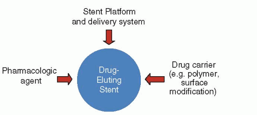
Figure 31.4 Components of drug-eluting stents.


Pharmacology

Following promising cell culture and in vitro development, the antirestenotic properties of a wide range of pharmacologic agents have been tested in humans (Figure 31.5). The two most clinically effective classes of agents have been the “rapamycin-analogue” (or “-limus”) family of drugs and paclitaxel. The principal mechanism of action of rapamycin (also known as sirolimus), and its analogues (including zotarolimus, everolimus, biolimus A9, novolimus, and amphilimus) is inhibition of the mammalian target of rapamycin (mTOR), which prevents cell cycle progression from the G1 to S phase.43 Two other rapamycin analogues that have been used on DES platforms—tacrolimus and pimecrolimus— have a different mechanism of action, binding directly to FK-binding protein (FKBP)-506 and thereby inhibiting the calcineurin receptor with downregulation of cytokines and inhibition of smooth muscle cell activity44; unlike the mTOR inhibitors, these agents have not been demonstrated to be beneficial. The other agent that has been used effectively in coronary DES (and more widely now in drug-eluting balloons and in peripheral DES applications) is paclitaxel. By interfering with microtubule function, paclitaxel has multifunctional antiproliferative and antiinflammatory properties, prevents smooth muscle migration, blocks cytokine and growth factor release and activity, interferes with secretory processes, is antiangiogenic, and impacts signal transduction.45, 46, 47 At low doses (similar to those in DES), paclitaxel affects the G0-G1 and G1-S phases (G1 arrest) resulting in cytostasis without cell death.45,48


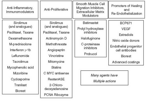
Figure 31.5 Potential antirestenotic agents for use with drug-eluting stents.


Polymers and Drug Delivery Systems

Early DES programs were plagued by the inability to predictably deliver a specific dose of active drug over the right time frame to the arterial wall.49 In order to more effectively regulate the dosing of antirestenotic agents, a drug carrier vehicle became necessary. In many respects, formulating and optimizing the drug carrier vehicle have proven even more complex than identification of the drug itself. Properties that must be considered for a controlled release vehicle include its biocompatibility, solubility, diffusivity and porosity, molecular size, weight and distribution, elongation, functional requirements, degradation products, durability, relative hydrophobicity, purity, availability, adhesion, crystallinity, sterilization, solvent solubility, biostability, miscibility, bioabsorbable versus permanent nature, evaporation rate, thermal properties, resistance to humidity and temperature extremes, compatibility with specific drugs, approval for implant use, processability (which relates to shelf life), and packaging requirements.

Numerous polymer-based drug delivery systems have since been developed, and are DES-specific (discussed below). While the polymer is instrumental in regulating the pharmacokinetics of drug delivery to the arterial wall (which is necessary for reduced neointimal hyperplasia), the polymer may also elicit deleterious vascular responses. Specifically, histopathologic studies have demonstrated hypersensitivity and eosinophilic inflammatory reactions and delayed endothelialization with first-generation DES that were not previously seen with BMS.50, 51, 52 Whether these vascular responses in humans are directly related to the polymer or to toxicity from the drug
itself is not well known, but in animal models these effects can be attenuated by modification of the polymer vehicle.53 It is believed that inflammation and delayed endothelialization play a role in the development of late stent malapposition, aneurysm formation, stent thrombosis and restenosis.50,54,55 For these reasons, there has been great interest in developing inert and biocompatible polymers, bioabsorbable/biodegradable polymers, and even polymer-free DES.


Generations of Drug-Eluting Stents

DES are often classified into several generations of development (Table 31.2). First-generation devices include the two DES that were initially approved for clinical use by most regulatory bodies, each of which utilized an early (currently outdated) BMS stent platform with early durable polymers (not specifically designed for biocompatibility) in order to deliver either sirolimus or paclitaxel. Second-generation devices (currently used in the vast majority of DES procedures) have incorporated more deliverable, thinner-strut stents with polymers that have been designed for biologic compatibility. Most second-generation DES utilize -limus (rapamycin) analogues. Future-generation DES will continue to undergo iteration, with further modifications of the base stent and use of biodegradable/bioabsorbable or polymer-free drug-carrier vehicles.


Table 31.2 Generational Classification of Drug-Eluting Stents


Generation


Drug


Polymer


Stent


First


Sirolimus or Paclitaxel


Not Specifically Designed for Biocompatibility


Early BMS Platforms


Cypher™


Sirolimus


Biostable mix of poly-n-butyl methacrylate and polyethylene-vinyl acetate


Bx Velocity™


TAXUS™ Express


Paclitaxel


Styrene-isobutylene-styrene (SIBS)


Express


TAXUS™ Liberté


Paclitaxel


Styrene-isobutylene-styrene (SIBS)


Libertéa


ION™ (TAXUS™ ELEMENT)


Paclitaxel


Styrene-isobutylene-styrene (SIBS)


Element (platinum-chromium)a


Second


Limus Analogues


Biocompatible Polymers


More Flexible, Thinner-Strut BMS


Endeavor™


Zotarolimus


Phosphorylcholine


Driver (cobalt alloy)


Xience V™ and Xience PRIME™


Everolimus


Vinylidene fluoride and hexafluoropropylene


Multi-Link Vision and Multi-Link 8 (cobalt-chromium)


Promus Element™


Everolimus


Vinylidene fluoride and hexafluoropropylene


Element (platinum-chromium)


Resolute™


Zotarolimus


Biolinx polymer


Integrity (cobalt alloy)


Biomatrix™


Biolimus A9


Abluminal poly-L-lactic acid (bioabsorbable)


Juno (stainless steel)


Nobori™


Biolimus A9


Abluminal poly-L-lactic acid (bioabsorbable)


S-stent


a Liberté and, in particular, Element BMS platforms are newer BMS platforms but are included in the first-generation due to the presence of the original TAXUS™ polymer.


FIRST GENERATION DRUG-ELUTING STENTS


The Cypher™ Sirolimus-Eluting Stent

The first DES to attain approval for human use was the Cypher™ stent (Cordis, Johnson and Johnson), with initial first-in-human studies as well as subsequent clinical trials
leading to its approval in Europe in 2002 and in the United States in 2003. Production of this stent was recently halted, but some description of the technology and initial studies that led to device approval is of historical interest, as the introduction of this stent ushered in the DES era of interventional cardiology. Sirolimus (rapamycin) is a highly lipophilic, naturally occurring macrocyclic lactone, which was first isolated from Streptomyces hygroscopicus found in a soil sample from Easter Island (also known as Rapa Nui) and was initially developed as an antifungal agent. Shortly thereafter, it became apparent that this agent also was a potent immunosuppressive, and was initially approved by the FDA (as Rapamune) for prevention of renal transplant rejection in 1999. The primary mechanism of action of inhibition of neointimal hyperplasia in sirolimus is thought to be related to its ability to bind to FKBP-12 in cells; the sirolimus-FKBP-12 complex then binds to and inhibits activation of mTOR, preventing progression in the cell cycle from the late G1 to S phase.43 Sirolimus has been demonstrated to have a marked effect on suppression of neointimal hyperplasia with low toxicity following sirolimus-eluting stent (SES) implantation in initial small and large animal studies.56,57

The stent platform for the Cypher™ SES was the Bx Velocity™ stent, a thick-strutted slotted tube with a closed cell design constructed from 316L stainless steel. The stent was coated with biostable (nonerodible) polymers consisting of poly-n-butyl methacrylate and polyethylene-vinyl acetate that are loaded with 140 µg/cm2 sirolimus. The slow-release (SR) formulation of the Cypher™ SES employed in clinical practice used a basecoat of blended polymers loaded with sirolimus as well as a topcoat of polymer alone (without sirolimus) that acted as a diffusion barrier, controlling the rate of drug release from the basecoat into the vessel wall. Approximately 80% of the sirolimus loaded on the stent was released within the first month after stent implantation.

In 1999, human experience with the Cypher™ SES was initiated at the Institute Dante Pazzanese of Cardiology in São Paulo, Brazil, and the Thoraxcenter, Rotterdam, The Netherlands, with the first-in-man (FIM) study in 45 patients with symptomatic de novo native coronary lesions <18 mm in length with RVD 3.0 to 3.5 mm. In this study, SES demonstrated marked suppression of neointimal hyperplasia measured by IVUS and quantitative coronary angiography at 4 months, and 1, 2, and 4 years.58 Serial angiography and IVUS have now been performed at 7 years, showing continued vessel patency without further late loss (Figure 31.6). These data led to the conduct of the larger RAVEL trial, in which 238 patients outside the United States with relatively simple de novo coronary lesions were randomized to either the Cypher™ SES or the uncoated Bx Velocity stent.36 The SES essentially eliminated late loss compared with BMS (mean of -0.01 mm versus 0.80 mm, P < 0.001), with a corresponding reduction in the rate of angiographic restenosis (0% versus 26%, P < 0.001).

On the basis of these data, the larger pivotal SIRIUS trial was conducted in the United States.59 SIRIUS was a 1058-patient randomized trial comparing the Cypher™ SES to the uncoated Bx Velocity in patients with vessel diameters
of 2.5 to 3.5 mm and lesion lengths of 15 to 30 mm. The primary endpoint, the rate of target vessel failure (TVF, a composite of cardiac death, myocardial infarction [MI], or target vessel revascularization [TVR]) at 9 months, was markedly lower among SES-treated patients (8.6% versus 21.0%, P < 0.001) (Figure 31.7). Additionally, SES resulted in a 60% to 80% relative reduction in composite adverse events in all examined subgroups in the trial. Among the 703 patients in whom 8-month routine angiographic follow-up was performed, mean in-stent late loss was markedly lower with SES (0.17 mm versus 1.00 mm, P < 0.001). By IVUS, the in-stent percent volumetric obstruction at 8 months was reduced from 33.4% with the Bx Velocity to 3.1% with the SES (P < 0.001), although late stent malapposition was present in 9.7% of Cypher™ SES patients versus 0% of Bx Velocity patients (P = 0.02).


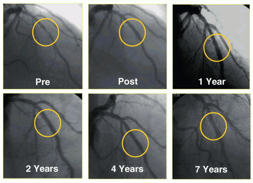
Figure 31.6 Seven-year follow-up of one of the initial sirolimus-eluting stent implantations from Institute Dante Pazzanese of Cardiology in São Paulo, Brazil, demonstrating sustained patency of the initially stented segment.

On the basis of these results, in April 2003 the Cypher™ SES became the first DES approved by the FDA, and this stent became one of the most studied devices in modern history, with a multitude of randomized trials and observational studies assessing its efficacy and safety. In their aggregate, these data demonstrate extremely low levels of in-stent late loss with SES (averaging ˜0.15 mm across studies), with an approximate 70% to 80% reduction in angiographic restenosis and clinical revascularization of the target lesion (TLR) compared to BMS. Longer-term follow-up with this device extending to 5 years and beyond has confirmed these findings. In these analyses, treatment with SES has resulted in sustained reductions in clinical restenosis endpoints with similar rates of death, MI, and stent thrombosis compared with BMS.60 In part due to the availability of newer stent platforms and designs, the manufacturer of this stent recently announced that the device would no longer be manufactured and sold, ending the stent’s tenure as the oldest DES in current clinical use.


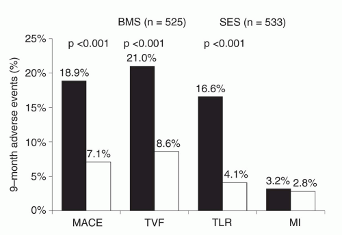
Figure 31.7 Primary results of the SIRIUS trial, the pivotal approval trial of the sirolimuseluting stent, demonstrating superiority of the sirolimus-eluting stent in reducing restenosis-related endpoints. SES, sirolimus-eluting stent; BMS, bare-metal stent; MACE, major adverse cardiac events; TVF, target vessel failure; TLR, target lesion revascularization; MI, myocardial infarction.


The Taxus™ Paclitaxel-Eluting Stent

The other first-generation DES that came to market soon after approval of SES was the TAXUS™ paclitaxel-eluting stent (PES). Paclitaxel, a highly lipophilic diterpenoid compound, was first isolated in 1963 from the pacific yew tree (Taxus brevifolius) and developed for its potent antineoplastic properties. Its principal action is to interfere with microtubule dynamics, preventing their depolymerization. This leads to widespread dose-dependent multicellular activity of the drug, including antiproliferative and antiinflammatory properties, reduced smooth muscle migration, blocking of cytokine and growth factor release and activity, interference with secretory processes, antiangiogenic effects, and impaired signal transduction.45, 46, 47 At low doses (similar to those in DES applications), paclitaxel affects the G0-G1 and G1-S phases (G1 arrest) resulting in cytostasis without cell death (probably via induction of p53/p21 tumor suppression genes).45,48 Systemic paclitaxel was shown to inhibit restenosis in a rat carotid injury model at levels more than 100-fold lower than that required for tumor cytotoxicity.46 Neointimal area was greatly reduced in a rabbit balloon-injury experiment using local paclitaxel administration,45 and stent-based paclitaxel elution from polymer-based systems has been shown to profoundly reduce intimal hyperplasia in rabbit iliac arteries for up to 6 months with dose-dependent efficacy and toxicity.61,62 The TAXUS™ PES (Boston Scientific, Natick, MA) consists of paclitaxel contained within a polyolefin derivative biostable polymer (styrene-isobutylene-styrene, referred to as SIBS (Translute™)), originally coated on the Nir stent and subsequently on the Express open-cell slotted tube stainless steel stent platform (PES(E), the device from which most of the randomized clinical trial data for this stent was derived). The base BMS has further evolved from the Express stent to the newer Liberté stent (PES(L), a more flexible, thinnerstrutted open-cell stainless steel slotted tube stent, and finally to a platinum-chromium Element stent. Depending on the relative ratio of paclitaxel to polymer, the stent may be formulated with varying release kinetics. The clinically available formulation of the TAXUS™ PES is the SR formulation, although the moderate-release (MR) formulation has also been tested in moderate-sized clinical trials. The SR stent has relatively more polymer to drug (paclitaxel concentration of 1 µg/mm2), with a coat thickness 18 µm, and approximately 8% in vivo paclitaxel elution in 30 days. The drug is eluted in a rapid burst phase over the initial 48 hours, followed by a slow, sustained release for the next 10 to 30 days, with the remainder sequestered in the bulk of the polymer matrix below the surface without pathways to the external environment (thus permanently retained on the stent).


The TAXUS clinical program evaluated the clinical safety and efficacy of the TAXUS™ PES in several clinical trials.63, 64, 65, 66, 67 TAXUS I and II evaluated the performance of the PES on the Nir stent platform in focal de novo disease, whereas TAXUS IV, V, and VI investigated the PES(E) stent in more complex lesions. All studies have used the SR formulation, except for one arm of the TAXUS II trial and TAXUS VI. Collectively, these trials demonstrate a marked decrease of binary restenosis with PES compared to BMS, with an approximate 60% to 75% reduction in the need for TLR, an effect that has been consistent across a range of patient and lesion subtypes. The study that ultimately led to device approval in the United States in 2004 was the TAXUS IV trial,65 which enrolled 1,314 patients with single de novo lesions with visually estimated lengths of 10 to 28 mm in native coronary arteries with an RVD of 2.5 to 3.75 mm. Patients were assigned to either a PES(E) stent or Express BMS control. The primary endpoint of TVR assessed at 9 months was reduced with the PES(E) from 12.0% to 4.7% (P < 0.001) (Figure 31.8). Follow-up angiography at 9 months demonstrated marked reductions in mean instent late loss (0.39 versus 0.92 mm, P < 0.001), and the rate of binary in-segment restenosis (7.9% versus 26.6%, P < 0.001). By IVUS, the in-stent percent volumetric obstruction at 8 months was reduced from 29.4% with the BMS to 12.2% with PES(E) (P < 0.001), and late stent malapposition at 9 months was present in 1.1% of PES(E) patients versus 2.2% of BMS patients (P = 0.62).

The PES(E) has been studied in numerous randomized trials and observational analyses, across a range of patient indications and lesion subsets. These studies have demonstrated consistent reductions in measures of neointimal hyperplasia and resultant reductions in clinical restenosis endpoints compared with BMS. Longer-term follow-up with this device has extended to 5 years and beyond, confirming the sustained efficacy of this stent.65 In these analyses, treatment with PES has resulted in sustained reductions in clinical restenosis endpoints, with similar rates of death, MI, and stent thrombosis with PES and BMS. Additionally, a series of comparisons between the first two approved devices (SES and PES) was reported in order to determine whether superiority could be established for a particular DES. In summary, the totality of evidence appears to indicate similar performance of SES and PES in routine de novo coronary artery lesions, despite a lower amount of neointimal hyperplasia with SES as assessed by IVUS and angiography.68, 69, 70, 71 Given the greater degree of late loss suppression with the SES, it was hypothesized that in the highest restenotic risk patients and lesions, this stent would hold an advantage over PES. Without a large-scale adequately powered randomized trial, however, these potential benefits remain unproven.


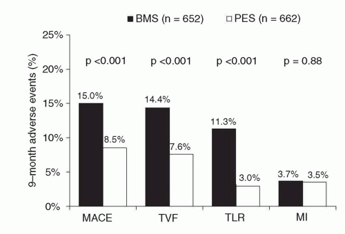
Figure 31.8 Primary results of the TAXUS-IV trial, the pivotal approval trial of the paclitaxel-eluting stent, demonstrating superiority of the paclitaxel-eluting stent in reducing restenosis-related endpoints. PES, paclitaxel-eluting stent; BMS, bare-metal stent; MACE, major adverse cardiac events; TVF, target vessel failure; TLR, target lesion revascularization; MI, myocardial infarction.

The commercially available PES has undergone several iterations, but is still generally considered a “firstgeneration” DES due to its use of an original polymer to elute paclitaxel. The PES(L) stent (using the same drug and polymer formulation as the PES(E), but with an improved stent platform) was approved for clinical use based upon the TAXUS ATLAS program, in which nonrandomized data from several PES(L) single-arm studies were compared to the treatment arms from prior TAXUS trials with the PES(E).72 More recently, the PES(L) has been replaced by the TAXUS™ Element stent (again, using the same drug and polymer formulation as the original TAXUS™ Express SR, but with an iterated stent platform using a platinum chromium alloy). The TAXUS™ Element stent (or ION stent) is the current commercially available version of PES in the United States. Approval of this stent required completion of the PERSEUS trial, which evaluated 1,262 patients with de novo “workhorse” atherosclerotic coronary lesions allocated in a 3:1 randomization to TAXUS™ Element versus PES(E).73 The TAXUS™ Element was demonstrated to be noninferior to PES(E) with respect to the primary endpoint of 12-month target lesion failure (TLF: 5.6% versus 6.1%, respectively) as well as the secondary endpoint of percentage diameter stenosis at 9-month angiographic follow-up (3.1% versus 3.1%, respectively). No differences in clinical outcomes were observed between the two randomized groups in this trial. The TAXUS™ Element stent has additionally been evaluated in smaller vessels in a prospective, single-arm trial comparing 224 patients treated with this stent with 125 lesion-matched historical Express BMS control subjects from the TAXUS V trial.74 In this analysis, the TAXUS™ Element was superior to the Express BMS with respect to late lumen loss (0.38 mm versus 0.80 mm, P < 0.001), and TLF (7.3% versus 19.5%, P < 0.001).


SECOND-GENERATION DRUG-ELUTING STENTS

Despite the demonstrated efficacy of first-generation SES and PES as observed in the initial and subsequent randomized trials of these devices, late reactions to first-generation DES polymers as well as delayed endothelialization and adverse vessel responses were described,54,75 potentially resulting in the most devastating complication of stent placement, namely late stent thrombosis. In order to mitigate some of the abnormal vessel responses associated with first-generation DES, several new devices have been introduced, with specific modifications implemented upon first-generation technology. These so-called second-generation DES (currently used in the majority of PCI) have incorporated more deliverable, thinnerstrut stents with polymers that have been specifically designed for biologic compatibility. Discussed below are clinical data relating to the most-studied second-generation devices, namely, everolimus-eluting stents (EES; Xience V/Promus and everolimus-eluting platinum chromium stent (Promus Element)); zotarolimus-eluting stents (ZES; Endeavor and Resolute); and biolimus A9-eluting stents (BES; Biomatrix).


Everolimus-Eluting Stents (Xience V™/Promus™)

In the EES (manufactured by Abbott Vascular (Santa Clara, CA) and distributed as the Xience V and now Xience PRIME stents, and also originally distributed by Boston Scientific as the Promus stent), everolimus (100 µg/cm2) is released from a thin (7.8 µm), nonadhesive, durable, biocompatible, fluorocopolymer consisting of vinylidene fluoride and hexafluoropropylene monomers, coated onto a low-profile (81 µm strut thickness), flexible, cobalt chromium stent. (The original Xience V base stent platform has been updated in the Xience PRIME stent to the Multi-link 8 BMS platform, a more deliverable version of the Vision platform). The release kinetics of EES are similar to that seen with sirolimus from the SES (˜80% of the drug released at 30 days, with none detectable after 120 days). The polymer is elastomeric, and experiences minimal bonding, webbing, or tearing upon expansion. Fluoropolymers have additionally been shown to resist platelet and thrombus deposition in blood-contact applications.21,76,77 The EES polymer has also been demonstrated to be nonin-flammatory in porcine experiments. Preclinical studies have demonstrated more rapid coverage of the stent struts with functional endothelialization with EES compared to SES, PES, or ZES.53

In the small SPIRIT First trial, the EES was shown to markedly reduce the extent of angiographic late loss at 6 and 12 months compared to the otherwise identical cobalt chromium Vision BMS.78 Subsequently, the EES has been studied in multiple randomized trials comparing this device to PES (the predominant comparator), SES, ZES, and BMS (Table 31.3).42,79, 80, 81, 82, 83, 84, 85, 86, 87, 88, 89 The large SPIRIT IV trial,42 enrolling 3,687 patients with stable coronary artery disease undergoing PCI of up to three lesions in three vessels, was a pivotal FDA approval study of the EES, randomizing patients to EES versus PES(E). While this study had broader inclusion criteria than first-generation DES approval studies, patients with unstable acute coronary syndromes, MI, thrombus, chronic occlusions, vein graft lesions, and true bifurcation lesions were excluded. The primary endpoint of TLF (a composite of cardiac death, target-vessel MI, or ischemia-driven TLR) was significantly lower at 1 year with EES compared to PES (3.9% versus 6.6%, P = 0.0008). Rates of stent thrombosis (0.3% versus 1.1%, P = 0.003), MI (1.9% versus 3.1%, P = 0.02), and TLR (2.3% versus 4.5%, P = 0.0008) were also lower with EES compared to PES. Longer-term follow-up of SPIRIT IV to 3 years85 has demonstrated sustained reductions in TLF, MI, and stent thrombosis with EES over PES (0.8% versus 1.9%), but narrowing of the initially observed difference in TLR with each stent (6.2% versus 7.8%, P = 0.06). However, both all-cause mortality (3.2% versus 5.1%, P = 0.02) and death or MI (5.9% versus 9.1%, P = 0.001) were reduced with EES compared to PES. These data from SPIRIT IV parallel results from the unrestricted “all-comer” COMPARE trial, in which 1,800 patients were randomized to EES versus PES(L). The primary endpoint of MACE at 1 year (death, MI, or TVR) was lower with EES compared to PES (6.2% versus 9.1%, P = 0.02), driven by reductions in stent thrombosis (0.7% versus 2.6%, P = 0.002), MI (2.8% versus 5.4%, P = 0.007), and TLR (1.7% versus 4.8%, P = 0.0002). Notably, between 1 and 3 years in this high-risk study cohort (in which only ˜15% of patients were maintained on dual antiplatelet therapy), fewer stent thrombosis, MI, and TLR events occurred with EES compared to PES.80

In contrast to the marked differences observed between EES and PES, smaller differences have been observed between EES and SES in several randomized trials. In the SORT OUT IV trial,87 2,774 unselected patients were randomized to EES versus SES and followed through the Danish Civil Registration System. Similar 9-month outcomes were observed between EES- and SES-treated patients although definite stent thrombosis occurred in fewer EES- than SES-treated patients at both 9 and 18 months (18 months: 0.2% versus 0.9%). In the 2,314-patient BASKET-PROVE multicenter trial comparing EES, SES, and BMS (the otherwise identical cobalt chromium Vision BMS) in large coronary arteries requiring >3.0 mm stents,93 EES, SES, and BMS were associated with similar rates of cardiac death or nonfatal MI at 2 years and the rate of TVR was similar between EES and SES. However, the rate of TVR was significantly lower with both EES and SES compared to BMS (3.1% for EES, 3.7% for SES, 8.9% for BMS), even in larger arteries with low rates of restenosis. The majority of comparative trials between EES and SES have demonstrated largely similar angiographic outcomes with EES and SES89,94,96 except for the ESSENCE-DIABETES trial,88 in which EES was associated with lower rates of angiographic late loss and binary restenosis in diabetic patients at 8 months compared to SES. Excepting this trial’s results, whether clinically apparent efficacy differences between EES and SES are manifest in the highest-risk patient and lesion subsets remains unknown.


Table 31.3 Randomized Controlled Trials of Everolimus-Eluting Stents


Trial Acronym and Reference


Study Cohort


EES Versus


Number Randomized (Planned Angiographic Follow-Up)


Latest Follow-Up to Date


Principal Findings


SPIRIT First78,90


Noncomplex CAD


BMS


60 (all)


5 y


EES versus BMS resulted in markedly reduced late loss and neointimal volume obstruction.


SPIRIT II82


Noncomplex CAD; up to 2 lesions


PES(E)


300 (all)


5 y


EES versus PES(E) resulted in lower 6-month angiographic in-stent late loss (0.11 ± 0.27 mm versus 0.36 ± 0.39 mm, P < 0.0001).


SPIRIT III83,91


Noncomplex CAD; up to 2 lesions


PES(E)


1,002 (564)


5 y


EES versus PES(E) resulted in lower 8-month angiographic in-segment late loss (0.14 ± 0.41 mm versus 0.28 ± 0.48 mm, P = 0.004), noninferior 9-mo rates of TVF (7.2% versus 9.0%, P = 0.31), and reduced rates of MACE at 1 y (5.9% versus 9.9%, P = 0.02) and 5 y (13.7% versus 20.2%, P = 0.007).


SPIRIT IV42,85


Noncomplex CAD; up to 3 lesions


PES(E)


3,687 (none)


3 y


EES versus PES(E) resulted in lower 1-y rates of TLF (3.9% versus 6.6%, P = 0.0008) and ischemia-driven TLR (2.3% versus 4.5%, P = 0.0008), with similar rates of cardiac death or target-vessel MI (2.2% versus 3.2%, P = 0.09). EES also resulted in lower rates of MI and stent thrombosis. At 3 y, these results were maintained although the difference in TLR was no longer significant (6.2% versus 7.8%, P = 0.06). 3-y mortality and death or MI were reduced with EES compared to PES (text).


COMPARE80,92


All-comers


PES(L)


1,800 (none)


3 y


EES versus PES(L) resulted in lower 1-y rates of the primary composite endpoint death, MI or TVR (6.2% versus 9.1%, P = 0.02). EES also resulted in lower rates of MI, stent thrombosis, and TLR (text). Between 1 and 3 y, EES resulted in less stent thrombosis, MI, and TLR events.


SPIRIT V Diabetes79


Diabetes


PES(L)


324 (all)


1 y


EES versus PES(L) resulted in lower 9-mo rates of angiographic in-stent late loss (0.19 ± 0.37 mm versus 0.39 ± 0.49 mm, P = 0.0001).


BASKET-PROVE93


Large coronary arteries (≥3.0 mm stents)


SES, BMS


2,314 (none)


2 y


EES and SES resulted in lower rates of TVR compared to BMS (3.1% and 3.7% respectively versus 8.9%). There were no differences between the three stent types in the rates of death, MI, or stent thrombosis at 2 y.


EXECUTIVE81


MVD, otherwise noncomplex CAD


PES(L)


200 (all)


9 mo


EES versus PES(L) resulted in lower 9-mo angiographic in-stent late loss (0.11 ± 0.27 mm versus 0.36 ± 0.39 mm, P = 0.008).


ISAR-TEST-486,94


Simple and complex CAD


SES


1,304 (all)


3 y


EES versus SES resulted in nonsignificantly different rates of in-segment late loss at 24 mo (0.29 ± 0.51 mm versus 0.31 ± 0.58 mm, P = 0.59). At 3 y, the rates of clinical outcomes were similar between EES and SES (for TLR: 12.8% versus 15.5%, P = 0.15).


SORT OUT IV87


Unselected patients


SES


2,774 (none)


18 mo


EES versus SES resulted in similar rates of the composite endpoint of death, MI, stent thrombosis, or clinically driven TVR at 9 and 18 mo (7.2% versus 7.6%, P = 0.64). Definite stent thrombosis at 18 mo was lower with EES (0.2% versus 0.9%, P = 0.03).


EXAMINATION95


STEMI


BMS


1,504 (none)


1 y


EES versus BMS resulted in similar rates of composite death, MI, or revascularization, but lower rates of TLR (2.2% versus 5.1%, P = 0.003). Definite/probable stent thrombosis at 1 y was lower in EES patients (0.9% versus 2.6%; P = 0.01).


EXCELLENT89


Noncomplex CAD


SES


1,443 (all)


9 mo


EES versus SES resulted in similar in-segment late loss at 9 mo (0.10 mm versus 0.05 mm, P for noninferiority = 0.02). Low rates of MACE were seen in both groups.


LONG-DES-III96


Long (≥25 mm) native coronary lesions


SES


450 (all)


9 m


EES versus SES resulted in higher in-segment late loss (0.17 mm versus 0.09 mm, P = 0.046), but similar instent late loss and in-stent binary restenosis as well as other clinical endpoints at 9 mo.


ESSENCE-DIABETES88


Diabetes


SES


300 (all)


1 y


EES versus SES resulted in


lower 8-mo angiographic in-segment late loss (mean 0.23 mm versus 0.37 mm, P = 0.02) and lower binary restenosis (0.9% versus 6.5%, P = 0.04). There were no differences in clinical outcomes between the two stents.


RESOLUTE


All-Comers84,97


Unselected


patients


ZES(R)


2,292 (460)


2 y


EES versus ZES(R) resulted in comparable 1-y rates of TLF (8.3% versus 8.2%, P = 0.92) and TLR (3.4% versus 3.9%, P = 0.50), although less definite stent thrombosis (0.3% versus 1.2%, P = 0.01) and definite/probable stent thrombosis (0.7% versus 1.6%, P = 0.05) were noted at 1 y At 2 y, similar rates of clinical endpoints were observed, with a trend toward less definite/probable stent thrombosis (1.0% versus 1.9%, P = 0.077).


TWENTE98


Unselected patients


ZES(R)


1,391 (none)


1 y


EES versus ZES(R) resulted in similar rates of TVR (8.1% versus 8.2%, P = 0.94) and other clinical endpoints including stent thrombosis at 1 year.


PLATINUM99


1 or 2 de novo native lesions


Pt-Cr EES


1,530 (none)


1 y


EES versus Pt-Cr EES resulted in similar rates of TLF (2.9% versus 3.4%, P = 0.60) as well as other clinical endpoints at 1 y.


EES, everolimus-eluting stents (Xience V/Promus); BMS, bare-metal stents; PES(E), paclitaxel-eluting stents (Taxus Express platform); PES(L), paclitaxeleluting stents (Taxus Liberté platform); ZES(R), zotarolimus-eluting stents (Resolute platform); Pt-Cr EES, platinum chromium EES; CAD, coronary artery disease; MVD, multivessel disease; MI, myocardial infarction; TLR, target lesion revascularization; TVR, target vessel revascularization; TLF, target lesion failure (cardiac death, target-vessel MI, or TLR); TVF, target vessel failure (cardiac death, MI, or TVR); MACE, major adverse cardiac events (cardiac death, MI, or TLR).


One intriguing attribute of EES that has emerged is the low rate of stent thrombosis observed with this stent. First demonstrated in SPIRIT IV and COMPARE, these findings have also been validated in several other studies, summarized in a metaanalysis of 13 randomized EES trials (N = 17,101) that demonstrated lower rates of ST with EES compared to non-EES DES.100 These data, combined with further observational validation of these findings,101 support the use of the second-generation EES over previously existing first-generation DES with respect to a safety advantage (in addition to efficacy). Further, whether EES can achieve lower or noninferior overall rates of stent thrombosis compared to BMS is an area of active interest, piqued by both preclinical data as well as studies such as the randomized EXAMINATION trial of 1,504 patients with ST-segment elevation myocardial infarction (STEMI),95 in which the rate of definite/probable stent thrombosis at 1 year was significantly lower in EES-treated patients compared to those treated with BMS (0.9% versus 2.6%; P = 0.01). Similarly, in a large network metaanalysis of head-to-head DES trials (49 trials, N = 50,844), the use of EES was associated with statistically significant reductions in 1- and 2-year stent thrombosis compared to other DES, as well as BMS.102 Whether EES can definitely reduce stent thrombosis compared to BMS is being actively tested in the randomized controlled HORIZONS-II trial.

Another iteration of EES has involved the use of everolimus eluted by the same stable fluropolymer as in the original EES, but on a platinum chromium stent platform (Promus Element, Boston Scientific, Natick, MA). This stent was evaluated in the randomized PLATINUM trial,99 which randomized 1,530 patients undergoing PCI of one or two de novo native lesions to treatment with the standard EES versus the Promus Element stent. The rates of efficacy and safety outcomes were very similar with both stents in this trial, which ultimately led to FDA approval of this EES platform.

In summary, in a broad cross-section of patients undergoing PCI, EES have shown significant improvements in safety and efficacy outcomes over first-generation DES. The finding of lower rates of stent thrombosis with EES, particularly compared to predecessor DES systems and in some cases even compared to BMS is notable, and suggests that this stent may have set a new standard for DES safety, if these findings can be further validated in larger adequately powered clinical trials.


Zotarolimus-Eluting Stents


Endeavor

Although studied contemporaneously with first-generation SES and PES, the zotarolimus-eluting Endeavor stent (ZES(E), Medtronic, Santa Rosa, CA) was originally conceived as a “second-generation DES,” rapidly eluting zotarolimus (10 µg per 1 mm stent length) from a thin layer (5.3 µm) of the biocompatible polymer phosphorylcholine from a flexible, low-profile (91 µm strut thickness) cobalt chromium stent. Phosphorylcholine is a naturally occurring phospholipid found in the membrane of red blood cells, and is resistant to platelet adhesion.103 The potencies of zotarolimus, everolimus, and sirolimus are roughly comparable, and zotarolimus is somewhat more lipophilic. However, the release rate of zotarolimus from Endeavor (˜90% within 7 days, 100% within 30 days) is significantly faster than everolimus and sirolimus are released from EES and SES stents respectively.

In the Endeavor I FIM study,104 ZES(E) was demonstrated to have a low rate of TLR (1%), despite a mean in-stent late lumen loss of 0.61 mm at 12 months. The ZES(E) was subsequently compared to its base BMS in the ENDEAVOR II randomized trial,105,106 conducted in 1,197 patients with noncomplex lesions. In this trial, ZES(E) was associated with lower rates of TVF and TLR at 9 months compared to BMS; these results were sustained at follow-up up to 5 years. Once again, 9-month angiographic in-stent late loss (at 0.61 mm) in this trial was greater than previously seen with either SES or PES, but compared to BMS, in-segment binary restenosis was reduced from 35.0% to 13.2% (P < 0.0001).

A series of head-to-head DES studies in the ENDEAVOR clinical trial program was launched with a 436-patient angiographic trial, ENDEAVOR III, which was designed to demonstrate noninferiority of ZES(E) to the Cypher SES. In this trial, the amounts of late loss and rate of restenosis at angiographic follow-up were significantly greater with ZES(E) compared to SES.107 Despite these findings, the overall rates of clinical restenosis endpoints were not dissimilar between treatment arms in this trial, and as such, the larger ENDEAVOR IV trial (N = 1,548) was conducted with a primary clinical endpoint (rather than an angiographic one). In this trial, which randomized patients with noncomplex coronary lesions to treatment with ZES(E) versus PES, despite greater late loss and angiographic restenosis with ZES(E) compared to PES, ZES(E) had noninferior 9-month rates of TVF and comparable 12-month rates of TLR.41 Rates of TLR were lowest and in fact indistinguishable between both stents particularly among patients who were assigned to receive clinical follow-up alone (rather than routine angiographic follow-up) (Figure 31.9), emphasizing a somewhat “artificial” clinical trial phenomenon previously described as the “oculostenotic reflex”.108 The ENDEAVOR IV findings ultimately led to device approval of ZES(E) in the United States. The 5-year follow-up of this trial has been recently presented,109 demonstrating comparable rates of TLR for ZES(E) compared with PES (7.7% versus 8.6%; P = 0.70). More notably, the ZES(E) demonstrated a superior late safety profile with lower very late stent thrombosis (0.4% versus 1.8%; P = 0.012) and a lower overall incidence of cardiac death or MI (6.4% versus 9.1%; P = 0.048) compared to PES at 5 years.

Several trials have compared ZES(E) to other DES in unrestricted patient populations. In the SORT OUT III trial,110 a trial notable for a design that employed follow-up through a nationwide clinical registry in Denmark, 2,333 patients
(nearly 50% of whom presented with acute coronary syndromes) were randomized to ZES(E) versus SES. In this trial, treatment with ZES(E) was associated with higher rates of 9-month major adverse cardiac events (MACE: cardiac death, MI, or TVR: 6% versus 3%, P = 0.0002), as well as endpoints of MI, stent thrombosis, and TLR, differences which persisted at 18 months (with the exception of stent thrombosis). The ISAR-TEST-2 trial was a three-way 1:1:1 randomized trial in 1,007 patients of an investigational combination sirolimus/probucol-eluting stent versus ZES(E) versus SES.111,112 Compared to SES, ZES(E) resulted in higher rates of late loss, angiographic restenosis (the primary endpoint), and TLR at 6 to 8 months, with similar rates of death, MI, and stent thrombosis. A larger study, the ZEST trial, randomized 2,645 patients with simple and complex coronary artery disease to ZES(E), SES, or PES.113,114 In this trial, while SES demonstrated the lowest degree of late loss and binary restenosis, ZES(E) was intermediate between SES and PES with respect to rates of MACE, TVR, and TLR. There were no significant differences in the 2-year rates of death, MI, or stent thrombosis between the two stents.


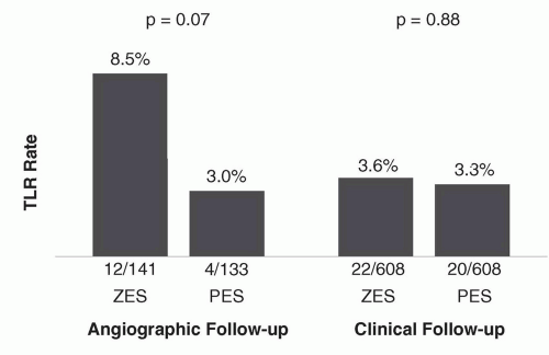
Figure 31.9 Rates of target lesion revascularization in the ENDEAVOR IV trial according to the performance of angiographic follow-up. The differences between stents are minimized among the majority of patients undergoing clinical follow-up alone. TLR, target lesion revascularization; ZES, zotarolimus-eluting stent; BMS, bare-metal stent.

Overall, both the pivotal approval trials within the ENDEAVOR clinical program as well as the investigator-initiated clinical trials of ZES(E) demonstrate lesser neointimal suppression with this stent compared to either SES or PES, resulting in lesser performance of this stent with respect to angiographically measured trial endpoints. However, ZES(E) is clearly superior in efficacy to BMS, and likely comparable to other stent platforms in reducing clinical restenosis in less complex lesions, particularly in the absence of routine angiographic follow-up. The findings of very low rates of late adverse safety events including very late stent thrombosis as well as cardiac death or MI115 with ZES(E) is a notable positive attribute of this stent, particularly in light of the potential ongoing thrombotic risks of SES and PES.116 In this regard the large, randomized PROTECT trial has completed enrollment of 8,800 patients to ZES(E) versus SES, and is the first clinical DES study powered to demonstrate a difference in stent thrombosis between two stent platforms (with ascertainment of the primary endpoint at 3 years).


Resolute

The Resolute stent (Medtronic Inc.) is similar to the Endeavor stent in that zotarolimus is eluted from the thin-strut cobaltalloy BMS platform (in this case, the updated and more deliverable Integrity cobalt-alloy BMS). However, instead of the phosphorylcholine coating of the Endeavor stent, the Resolute stent employs the BioLinx tripolymer coating, consisting of a hydrophilic endoluminal component and a hydrophobic component adjacent to the metal stent surface. This polymer serves to slow the elution of zotarolimus, such that 60% of the drug is eluted by 30 days and 100% by 180 days, making this the slowest rapamycin analogue-eluting DES.

In the single-arm RESOLUTE trial, ZES(R),117 the primary endpoint of in-stent late lumen loss at 9-months was 0.22 mm, and the in-segment binary restenosis rate was 2.1%, both significantly less than seen with other studies of ZES(E) or BMS. Low rates of MACE, TLR, and ARC definite/probable stent thrombosis were observed. Two-year data from this study have demonstrated TLR, TVR, and TVF rates of 1.4%, 1.4%, and 7.9%, respectively, with no late stent thrombosis events.118

The large RESOLUTE All-Comers randomized trial of ZES(R) versus EES was conducted in 2,292 patients84; this trial sought to enroll a more unselected patient population than in prior pivotal DES trials. The rate of the primary endpoint of TLF at 1 year was similar to ZES(R) and EES (8.2% versus 8.3%, P for noninferiority < 0.001). In this trial, the rates of death, cardiac death, MI, and TLR were similar with both stents, but both definite and definite or probable stent thrombosis occurred less frequently with EES at 1 year. Insegment late loss at 13 months (after ascertainment of the primary clinical endpoints) was slightly greater with ZES(R) compared to EES (0.15 mm versus 0.06 mm, P = 0.04), but there were no differences in rates of binary restenosis among the 460 patients undergoing angiographic follow-up. At 2 years, similar rates of clinical endpoints including TLF, TVF, MI, TLR, and TVR were observed, with a trend toward less stent thrombosis with EES (1.0% versus 1.9%, P = 0.077), predominantly driven by events within the first year.97 Three patients in each group (0.3%) had very late stent thrombosis (thrombosis occurring beyond 1 year). One additional investigator-initiated randomized trial of ZES(R) and EES has been reported; in the TWENTE trial98 1,391 unselected patients were randomized between these two stents. Notably, “off-label” indications occurred in >75% of patients enrolled. At 1 year, the primary endpoint of TVF was similar with both
stents (8.1% versus 8.2%, P = 0.94), with no observed differences in other clinical endpoints, including stent thrombosis (definite/probable: 0.9% for ZES(R) versus 1.2% for EES).

In summary, the ZES (Resolute platform) is the first stent to demonstrate comparable overall safety and efficacy to the EES, although slight differences in angiographic and clinical outcomes between these stent platforms may exist. Larger studies and longer-term follow-up are required to assess whether these device-specific performance characteristics influence outcomes in actual clinical practice, and whether the long-term safety of this stent is maintained.


Biolimus A9-Eluting Stents (BioMatrix)

The BioMatrix (Biosensors International, Switzerland) stent (BES) elutes biolimus A9 (concentration 15.6 µg/mm), a semisynthetic rapamycin analogue with similar potency but greater lipophilicity than sirolimus, from a stainless steel platform. The stent platform that originally was the S-stent is currently the Juno BMS platform, in the BioMatrix Flex iteration of BES. Of note, the Nobori DES (Terumo Medical Corporation, Japan) is a similar BES that releases biolimus using the same polymer system with a different BMS platform. The Nobori DES has demonstrated favorable results compared to PES and SES in three modest-sized randomized trials.119, 120, 121 BES are unique, especially compared to the previously described firstand second-generation DES, in that biolimus A9 is eluted from PLLA, a biodegradable polymer which is applied solely to the abluminal stent surface. The biolimus A9 and PLLA are coreleased, and the polymer is converted via the Krebs cycle into carbon dioxide and water after a 6- to 9-month period. Conceptually, such a stent might not be prone to late inflammatory reactions as are occasionally seen with durable polymers, and thus result in improved outcomes after 1 year.

The BioMatrix BES was first tested in the randomized STEALTH trial in which 120 patients with single de novo coronary lesions received either a BES or a bare-metal S-stent.122 Treatment with BES resulted in lower in-stent late loss at 6 months (0.26 mm versus 0.74 mm for BMS, P < 0.001). The largest trial examining the safety and efficacy of BES was the LEADERS trial, which randomized 1,707 “all-comer” patients (55% of whom had acute coronary syndromes) to BES versus SES.123 Similar rates of all clinical endpoints were observed at 9 months with both BES and SES, including the primary study endpoint, which was the composite of cardiac death, MI, or TVR (9.2% versus 10.5%, P = 0.39). Among the 427 patients allocated to angiographic follow-up at 9 months, in-stent late loss and binary restenosis were similar with both stents. Longer-term follow-up of LEADERS to 4 years has been recently reported (Figure 31.10).124 Over the entire follow-up period, the rate of the composite primary endpoint of cardiac death, MI, or clinically indicated TVR was lower with BES compared to SES (19% versus 23%, P = 0.039), with gradual separation of respective event curves over time. Additionally, while overall definite/probable stent thrombosis rates were not significantly different (3% for BES versus 5% for SES, P = 0.20), the rate of very late definite/probable stent thrombosis was significantly lower with BES (6 events (1%) versus 20 events (2%), P = 0.005). Similar results were observed when assessing the endpoint of definite stent thrombosis.

Collectively, these data demonstrate that BES has similar efficacy as the first-generation devices, with a favorable safety profile that emerges particularly beyond 1 year. However, much larger and adequately powered studies will be required to determine whether BES, or other devices with bioabsorbable polymers, offer true and sustained clinical advantages to the best-in-class second-generation DES with durable polymers. Several studies investigating these hypotheses are ongoing.


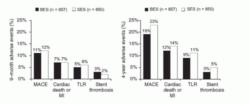
Figure 31.10 Principal clinical endpoints at 1 year (left) and 4 years (right) from the randomized all-comers LEADERS trial of a biolimus A9-eluting stent compared to a sirolimus-eluting stent. BES, biolimus A9-eluting stent; SES, sirolimus-eluting stent; MACE (major adverse cardiac events) denotes cardiac death, myocardial infarction (MI), or clinically indicated target vessel revascularization; stent thrombosis refers to Academic Research Consortium (ARC) definite or probable events.


CONCERNS REGARDING SAFETY OF DRUG-ELUTING STENTS AND POOLED COMPARISONS OF DRUG-ELUTING STENTS AND BARE-METAL STENTS

The evidence base for initial DES approvals by the FDA consisted primarily of randomized controlled trials enrolling largely stable patients with relatively noncomplex, single, de novo coronary artery lesions. Data from these early studies demonstrated similar rates of death and MI among DES and BMS-treated patients.39,125 Yet, due to their potent efficacy, DES are used “off label” (in higher-risk patients and in more complex lesions) in 60% to 70% of cases,126 leading to concerns about the safety and appropriateness of the routine use of DES in the “real world.” Moreover, most randomized studies (especially those conducted early in the DES era) included primary outcomes of interest that focused upon stent efficacy, rather than absolute safety. As such, evidence of the safety of DES has come from two sources—randomized controlled trials, which are usually small to modest in size, and typically underpowered to assess safety endpoints such as death, MI, and stent thrombosis, as well as large-scale observational studies, which provide a broader look at the real-world use of DES and allow more generalizability and power.


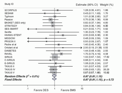
Figure 31.11 Mortality in randomized trials comparing drug-eluting stents to bare-metal stents (from Kirtane et al., Circulation 2009), demonstrating similar overall mortality of both stent types. DES, drug-eluting stent; BMS, bare-metal stent.

A number of analyses have amalgamated trial data across clinical studies to increase overall sample size. In particular, these studies have attempted to address one of the prominent limitations of individual DES studies, namely the limited power to detect differences in low-frequency safety endpoints. In the largest and most comprehensive metaanalysis of first-generation DES versus BMS studies (including 9,470 patients from 22 randomized trials and 182,901 patients from 34 observational studies), the use of DES in randomized trials was associated with comparable rates of mortality and MI, with a 55% relative reduction in TVR (Figure 31.11).39 In the observational studies included separately in this analysis (Figure 31.12), significant heterogeneity was observed, and treatment with DES was in fact associated with significant reductions in overall death, MI, as well as TVR. The differences observed between the findings of randomized trials and observational studies included in this analysis highlight the difficulty in assessing nonrandomized active treatment comparisons through an observational study design. In another metaanalysis, Stettler and colleagues incorporated comparative data from SES versus BMS trials, PES versus BMS trials, and SES versus PES trials in a statistical “network” of trials to discern treatment effects across all
included trials.127 In this analysis of 38 trials including data from 18,023 patients, TLR was lower with SES and PES compared to BMS, with similar mortality among patients treated with SES, PES, and BMS. In this analysis, a reduction in the hazard of MI was observed with SES compared to both BMS (hazard ratio [HR] 0.81, 95% credibility interval 0.66 to 0.97, P = 0.030) and PES (HR 0.83, 0.71 to 1.00, P = 0.045).


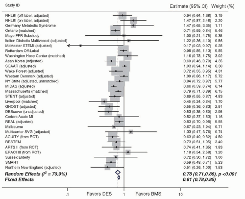
Figure 31.12 Mortality in observational studies comparing drug-eluting stents to bare-metal stents (from Kirtane et al., Circulation 2009), demonstrating a reduction in mortality with drug-eluting stents. DES, drugeluting stent; BMS, bare-metal stent.

In addition to these and other analyses, numerous observational studies have focused upon the examination of lowfrequency safety endpoints when comparing first-generation DES to BMS, across a wide range of clinical indications. More than 50 nonrandomized comparisons between DES and BMS have been published and/or presented to date. Aside from the initial publication of data from SCAAR registry128 that was subsequently revised with the addition of longer term follow-up,129 the majority of these studies have demonstrated favorable safety for DES compared to BMS. For example, in the largest such analysis of DES safety, which was conducted using data from 262,700 Medicare beneficiaries in the United States, the use of DES was associated with lower rates of death (13.5% versus 16.5%, P < 0.001) and MI (7.5% versus 8.9%, P < 0.001) with minimal differences in bleeding.130

Only gold members can continue reading. Log In or Register to continue

Stay updated, free articles. Join our Telegram channel

Jun 26, 2016 | Posted by in CARDIOLOGY | Comments Off on Coronary Stenting

Full access? Get Clinical Tree

Get Clinical Tree app for offline access