Blood Gas Classification


Blood Gas Classification


INTRODUCTION


The techniques used in the acquisition of arterial blood have been discussed. In this chapter, the first step in the clinical application of arterial blood gases, arterial blood gas classification, is explored.


As described previously, the arterial blood gas report is the basis and cornerstone in the assessment and management of clinical oxygenation, ventilation, and acid-base disturbances. The initial objective in clinical management of these areas is to classify the blood gas information into one of several possible general categories, for example, “Partially Compensated Metabolic Acidosis with Mild Hypoxemia.”


Sometimes this “naming” of the blood gas is referred to as blood gas interpretation. Notwithstanding, simply stating that a patient has a metabolic acidosis without understanding the nature of the acid-base disturbance is taking a very narrow view of “interpretation” and does not facilitate optimal patient treatment. Therefore, in this text, “interpretation” is considered in a much broader context to include detailed analysis and evaluation of the blood gas in conjunction with other patient clinical and laboratory information. This requires critical (diagnostic and therapeutic) thinking and is, indeed, our goal in the clinical management of acid-base balance, ventilation, and oxygenation.


To accomplish these important tasks, the first important step in interpretation is “blood gas classification.” Even the novice clinician dealing with critical care patients must learn immediately how to “classify an ABG” correctly and expeditiously. Likewise, even the novice must be able to quickly identify critical values and life-threatening situations. These objectives are the aim of this chapter.


SYSTEMATIC APPROACH


The vital nature of blood gas information requires a careful, thoughtful approach. The variety of data that may be reported (e.g., [HCO3], SaO2, PaO2, [BE]) is a potential source of confusion for the novice. Therefore, it is important to process the data on a blood gas report in an orderly, systematic, and thorough manner. A step-by-step approach ensures reproducible results and helps to avoid confusion and omissions.


Although acid-base balance, ventilation, and oxygenation status can often present interrelated problems and issues, individual and separate evaluations of these areas sometimes help to focus and clarify thinking. Although the sequence is somewhat arbitrary, in the classification system presented here, acid-base classification (which also includes ventilation evaluation) is presented first and is followed by classification of blood oxygen levels.


ACID-BASE STATUS


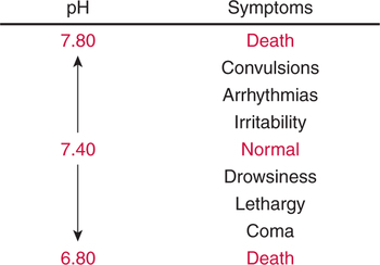
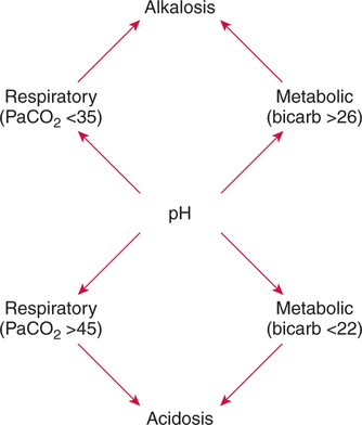
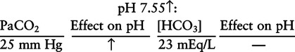
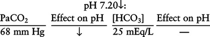
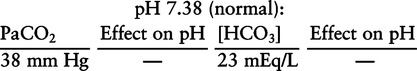
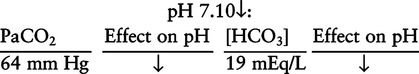
There are three steps in this simple ABC approach to acid-base classification; these steps are shown in Box 2-1. First, Acid-base status (overall body conditions) is assessed by classifying the arterial pH. The arterial pH is the single best index of composite acid-base status in the body.


Second, the Basic primary problem(s) (general type of acid-base disturbance) present is characterized as either respiratory, metabolic, or both. In this step, we attempt to identify the primary general abnormality that is (are) tending to pull the pH away from normal.


Next, Compensation is assessed. The body’s normal response to a single, primary, acid-base disturbance is to alter the acid-base component not primarily affected (respiratory [PaCO2] or metabolic ([HCO3]) in the opposite direction of the primary problem. This secondary acid-base change is referred to as compensation. Because this is an expected occurrence in normal individuals, each blood gas should be evaluated and classified according to the presence and extent of acid-base compensation.


Regarding the sequence of specific indices to be analyzed on a blood gas report, it is recommended that the pH be evaluated first as an index of overall Acid-base balance (Box 2-2). The PaCO2 (respiratory status) is evaluated next followed by [HCO3] (metabolic) evaluation. Assessment of both of these indices is necessary to complete both step 2 (Basic primary problem determination) and step 3 (Compensation assessment).


As we will see later in this chapter, the clinician should then analyze the PaO2 (oxygenation classification) and consider it in respect to the FIO2 (i.e., the concentration of oxygen being inspired) and lung oxygen exchange efficiency. Again, Box 2-2 summarizes the sequence of indices to be evaluated.


pH Assessment


Clinical Significance


The pH reported on an arterial blood gas sample is the single best index of overall acid-base status in the body. It is a composite reflection of the net interaction of all acids, bases, buffers, and compensatory mechanisms. It is the logical starting point in comprehensive acid-base assessment. Furthermore, if the pH is severely disturbed and the patient is in a life-threatening situation, this can be identified immediately.


The arterial pH is measured in the blood plasma and reflects quantitatively the hydrogen ion activity in this extracellular fluid compartment. Although the extracellular pH is not identical to the important intracellular pH, alterations in the two values tend to move in similar directions and correlate closely. Thus, the pH on a blood gas report is a good indicator of overall acid-base conditions.


Clinical Manifestations of Abnormal pH


Seemingly slight alterations in blood pH may have profound, life-threatening effects on body chemistry. This is likely due to the small size of the hydrogen ion and the vast number of chemical reactions it may impact. Clinically, it is useful to be aware of the typical manifestations associated with a particular pH disturbance.


Low arterial pH has a generalized depressive effect on the human nervous system (Fig. 2-1).173 Symptoms may include drowsiness and lethargy. Regardless of the precipitating cause, a very low pH (i.e., pH <7.10) is often associated with coma. A pH of less than 6.80 for any extended period is generally considered to be incompatible with life.


A high blood pH, on the other hand, has a general tendency to excite the central nervous system. Irritability or tetany may be manifest. When the heart muscle becomes more irritable, serious arrhythmias (abnormal beats) may result. When pH remains very high, convulsions may occur. A pH greater than 7.80 is generally considered to be incompatible with life. The clinician should always take note of the patient’s symptoms to substantiate, refute, or clarify laboratory findings and disease.


Classification of pH


BASIC (PRIMARY) ACID-BASE DISTURBANCE(S)


It has become standard diagnostic practice to characterize acid-base pathologic conditions into one of two general categories: respiratory and/or metabolic problems. This basic pathologic condition, sometimes referred to as a primary acid-base disturbance, is the root cause or problem that has led to (or has an inclination or tendency to lead to) overall acid-base disruption.


To determine the primary problem, the clinician should first evaluate (or classify) the respiratory status using the PaCO2. This is followed by evaluation of the metabolic (i.e., non-respiratory) acid-base status using either the plasma bicarbonate concentration [HCO3] or the base excess concentration [BE]. By understanding how these two indices may alter pH, the primary problem or problems can be easily identified as described subsequently.


Respiratory Acid-Base Status


Evaluation or classification of the respiratory component of acid-base balance is a logical starting point in “basic primary problem” determination because the respiratory system is the major organ system responsible for acid excretion and the moment-to-moment regulation of pH.


Regarding acid-base balance, the specific role of the lungs is to excrete carbonic acid at exactly the same rate at which it is being produced by the tissues as a result of carbon dioxide production via metabolism. Therefore, if the lungs are properly excreting carbonic acid, blood leaving the lungs (i.e., arterial blood) should have a constant, normal level of carbonic acid.


Measurement of the carbonic acid levels in the blood leaving the lungs would thus provide us with an index of lung effectiveness in acid-base balance (i.e., carbonic acid excretion). High carbonic acid levels in the arterial blood would indicate that the lungs are failing to adequately excrete this acid. Conversely, low carbonic acid levels in the blood would indicate excessive excretion and depletion of the body stores. Because arterial blood is a mixture of all the blood that has just left the lungs, it is ideal for this assessment.


The technical problem with this approach to respiratory acid-base assessment is that carbonic acid levels in the blood are very low, and measurement of carbonic acid levels is not technically feasible. Fortunately, however, there is a direct linear relationship between arterial carbonic acid levels and PaCO2. Thus, when PaCO2 is increased, it is indicative of increased carbonic acid in the blood, which tends to decrease pH (acidosis).


Logically, the converse is also true. A decreased PaCO2 indicates below-normal levels of carbonic acid in the arterial blood, which tends to elevate pH (i.e., alkalosis). In summary, the adequacy of carbonic acid excretion (respiratory acid-base function) can be assessed simply by evaluating PaCO2.


PaCO2 Classification


A normal level of carbonic acid in the arterial blood corresponds to a PaCO2 level of 35 to 45 mm Hg, which is shown in Table 2-2. Indeed, PaCO2 can be used simply to evaluate and classify the respiratory acid-base status.


Inverse Relationship (PaCO2–pH)


To classify arterial blood gases correctly, it is crucial to understand that the relationship between PaCO2 and pH is inverse. In other words, when PaCO2 is high (i.e., respiratory acidosis), pH will tend to decrease due to the accumulation of carbonic acid. Conversely, when PaCO2 is low (i.e., respiratory alkalosis), pH will tend to increase due to the depletion of carbonic acid stores in the blood.


A very common classification error for the novice is to see an abnormal pH and PaCO2 and assume the pH has changed because of the PaCO2, despite the fact that the change in pH and PaCO2 is not inverse.


For example, if pH is 7.20 (acidosis) and PaCO2 is 20 mm Hg (respiratory alkalosis), both of these values are abnormal; however, the change in pH cannot be due to the respiratory condition (i.e., this is not a primary respiratory acid-base problem) but must be due to some non-respiratory condition (i.e., metabolic acidosis).


If indeed the abnormal PaCO2 had caused an abnormal pH, the pH would be high (alkalosis) because the relationship must be inverse to assume cause and effect. Therefore, in this example, it is clear that the change in pH must be due to some other (non-respiratory) cause. To reiterate this important principle; if the change in pH is due to a respiratory condition, the relationship between pH and PaCO2 must be inverse.


Metabolic Acid-Base Status


All the numerous conditions that may potentially alter pH have been grouped into two major categories to facilitate differential diagnosis. Blood gas acid-base evaluation involves classification of the data based on these two components. Respiratory disturbances include all those conditions that alter PaCO2 levels in the blood. Metabolic disturbances, on the other hand, are defined by exclusion. Any acid-base disturbance that is not respiratory in origin is called a metabolic disturbance.


In some cases, the adjective metabolic may actually be misleading because many non-respiratory acid-base disturbances (e.g., vomiting) do not involve changes in “metabolism.” In fact, some authors have suggested that the adjective metabolic should be replaced with the adjective non-respiratory.174 Nevertheless, the term metabolic is well ingrained in clinical medicine and is used throughout this text.


The PaCO2 is a specific, reliable, accurate, and simple indicator of respiratory acid-base disturbance. No single metabolic acid-base index perfectly fits this description. In some cases, metabolic indices will deviate artifactually from their normal ranges owing to reasons unrelated to primary acid-base disorders. Nevertheless, a change in the numerical value of a metabolic index is most commonly due to a metabolic acid-base disturbance and the novice should assume this to be the case. In later chapters, exceptions to this rule will be discussed.


[HCO3] Classification


Although various different metabolic indices have been advocated through the years, the plasma bicarbonate concentration [HCO3] (sometimes referred to as the actual bicarbonate) is probably the most widely used index and is seen on many clinical, professional, credentialling examinations. Initial blood gas classification examples in this text will include only the [HCO3] as a metabolic index to avoid confusion. Also, as mentioned previously, there are some uncommon situations when bicarbonate values may be misleading (discussed in Chapter 5). It is best for the novice to assume the bicarbonate is always a clear and concise indicator of metabolic status. Logically, one should first learn the general rules of classification and later address unusual exceptions.


Identification of Primary Acid-Base Disturbances


As previously described, the pH should be evaluated first to get an overall view of acid-base status. Then, the PaCO2 and [HCO3] should be evaluated. If all three parameters (pH, PaCO2, [HCO3]) are in their normal ranges (Example 2-1), the classification is simply Normal Acid-Base Status and therefore no primary acid-base problem is present.


Example 2-1

Classification of Primary Blood Gas Problems


image


When pH is outside the normal range, one must determine the basic (primary) acid-base disturbance. The key to successful classification is to identify whether the primary problem is respiratory (PaCO2 is abnormal and inverse direction to pH change) or metabolic ([HCO3] is abnormal and change is in the same direction as pH).


Table 2-4 highlights these key relationships, which must be memorized to identify the primary acid-base problem. Figure 2-2 illustrates the paths by which one can identify primary acid-base disturbances in patients with acidosis or alkalosis. In some less common cases, both respiratory and metabolic components may be pulling pH in the same direction. When there are two primary acid-base problems both pulling pH in the same direction, the problem is said to be “Mixed” or “Combined.”


In Example 2-2, the primary problem is clearly metabolic because [HCO3] is decreased (normal 24 ± 2 mEq/L) along with a decreased pH (direct relationship). PaCO2 is within the normal range so this cannot be a primary respiratory acid-base problem. This would be termed a metabolic acidosis.


Example 2-3

Classification of Primary Blood Gas Problems


image


Example 2-4 shows a respiratory and a metabolic acidosis. This blood gas actually demonstrates two primary problems both pulling the pH in the same direction (lower). Again, PaCO2 is increased (which tends to lower pH because the relationship is inverse) and [HCO3] is decreased (which tends to lower pH because the relationship is direct). Thus, PaCO2 indicates a respiratory acidosis and [HCO3] indicates a metabolic acidosis. This may also be classified as a mixed or combined acidosis.


A blood gas such as that in Example 2-4 may accompany cardiac arrest. The respiratory acidosis results from decreased ventilation, whereas the metabolic acidosis is a result of anaerobic (without oxygen) metabolism and lactic acid accumulation. Although somewhat inter-related, two primary and separate basic acid-base problems coexist.


Example 2-4

Classification of Primary Blood Gas Problems


image


Example 2-5 is one of the most common types of blood gases seen and also one of the most commonly misclassified, particularly by the novice. As previously stated, many inexperienced clinicians, after looking at pH and PaCO2, incorrectly classify this as simply respiratory alkalosis. The logic is that pH and PaCO2 are both abnormal; so the abnormal respiratory condition must be the cause of the overall pH acid-base disturbance. The error in this logic is failing to remember the relationship between PaCO2 and pH as shown in Table 2-4. Whenever the individual has a primary respiratory acid-base disturbance, the relationship between PaCO2 and pH must be inverse.


This basic primary acid-base disturbance in this blood gas is a metabolic acidosis because a low [HCO3] tends to lower pH and indeed that explains the abnormally low pH. The low PaCO2 (respiratory alkalosis that tends to increase pH) is actually due to the normal compensatory response of the body, which is described later in this chapter. In summary, when determining primary problems, the key is to understand the relationships in Table 2-4.


Base Excess [BE] Assessment


An alternative metabolic index with fairly widespread popularity is the base excess [BE]. It is noteworthy for the novice in arterial blood gas classification that the base excess and plasma bicarbonate both provide the clinician with the same general clinical information regarding acid-base balance. There is, in fact, no need to evaluate both of these indices.


For the novice, it is recommended that, given a particular blood gas, only one of the metabolic indices should be classified to avoid confusion. Some unusual situations are discussed later in Chapter 5, when the two indices may not completely agree with each other.


If both indices are reported at your institution, the index most commonly used in your particular institution or region should be used for classification. Beginning exercises in this text will use bicarbonate as the metabolic acid-base index. In later examples, the base excess or both metabolic indices (i.e., [HCO3] and [BE]) values may be given.


From a numerical standpoint, the [BE] is easier to understand than bicarbonate since the normal value for base excess is 0 ± 2 mEq/L. A base excess in the negative range (metabolic acidosis) is sometimes called a base deficit. Nevertheless, even in the presence of a “base deficit,” this index is still usually referred to as the base excess.


Obviously, if you are using [BE] as the metabolic index, the same rules apply. An elevated [BE] (e.g., +5 mEq/L) would increase pH and would be termed a metabolic alkalosis (see Table 2-3). A lower [BE] (e.g., -5 mEq/L) tends to lower pH and would be termed a metabolic acidosis.


COMPENSATION ASSESSMENT


Compensation is defined as return of an abnormal pH toward normal by the component (i.e., respiratory or metabolic) that was not primarily affected. For example, when an abnormal respiratory acidosis (i.e., primary pathologic respiratory acidosis) occurs, the body (specifically the kidneys) responds by developing a compensatory increase in blood base (i.e., secondary metabolic alkalosis). The compensatory response helps protect pH and prevents large, abrupt, dangerous swings in pH. Conversely, in the presence of an abnormal (primary) metabolic acidosis, the respiratory system reduces blood PaCO2 levels (i.e., secondary respiratory alkalosis) in an attempt to bring the pH toward normal.


Uncompensated Acid-Base Problems


When one of the acid-base components (i.e., respiratory or metabolic) is abnormal and the other is within the normal range, the abnormal condition is said to be uncompensated (e.g.,uncompensated respiratory acidosis). The absence of compensation may be seen in respiratory acid-base problems that have developed rapidly or when the body is unable to compensate due to disease or some other reason.


Uncompensated respiratory acid-base problems generally indicate that the problem is of recent origin (i.e., acute) and that the kidneys have not had a sufficient time to manifest measurable compensation. Renal compensation is a relatively slow process in that it takes the kidneys considerable time (i.e., 48 to 72 hours) to achieve maximal compensation. As a general rule, it is unusual and, indeed, signals another patient problem when an acid-base disturbance remains uncompensated for a long time.


Total classification of acid-base status in Example 2-6 would be uncompensated respiratory acidosis. The patient has respiratory acidosis as described in the previous section because PaCO2 and pH are inverse. Regarding compensation, the respiratory acidosis is uncompensated because no increase in [HCO3] (no secondary metabolic alkalosis) above the normal range is seen.


Stay updated, free articles. Join our Telegram channel

Jul 10, 2016 | Posted by in RESPIRATORY | Comments Off on Blood Gas Classification

Full access? Get Clinical Tree

Get Clinical Tree app for offline access