Fig. 3.2 Current flows between different areas of the heart either when some areas have depolarized and others are still to depolarize or, conversely, when some areas have repolarized and others are still to repolarize. (a) Here depolarized cells are shown with excess negative charge, which flows in the direction of the depolarization wavefront. (b) This shows the current flow loops round the dipole, producing current flow to the side and behind the depolarization wave.
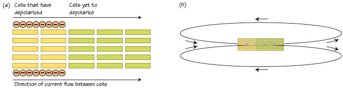
Fig. 3.3 This shows a more complex and so more realistic pattern of current flow than Fig. 3.2b. In the centre is a dipole (labelled as – or + for the polarity at either end), which generates a complex series of looping currents around itself.
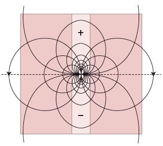
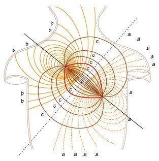
Fig. 3.4 The surface correlates of internal current flow, projected onto a torso. The pattern of current flow is complex. There is no current flow towards an observing electrode at right angles to the dipole, and maximal flow directly in front or behind the dipole.
The basis of the ECG
The ECG is a clever device designed to detect current flow. The heart generates electricity, which is transmitted to the chest wall, where it can be detected. The ECG records the pattern of spread of electricity in the various phases of the cardiac cycle. Its utility relies on the pattern of spread changing in a characteristic fashion in many diseases. In understanding the ECG, be aware that:
- Electrons carry current, which flows from areas with negative charge to areas with more positive charge; when current moves towards an observing electrode a positive deflection results and vice versa. The ECG only shows a deflection when current is moving in the heart. No current flow means no ECG deflection.
- The basis of current flow around the heart starts off as current flow within individual heart cells, which induce current flow between cells. With depolarization positively charged Na+ ions move into the cell; with repolarization, positively charged K+ move out of cells – this leaves excess negative charge outside the cell at the start of the cardiac cycle, excess positive charge outside the cell at the end of the cycle.
- Current flows from areas just depolarized (excess extracellular negative charge) into areas yet to depolarize, then onto an observing electrode. This current flow depolarizes neighbouring cells, firing action potentials, and in sequence fully depolarizing the whole heart. When fully depolarized, the extracellular charge throughout the heart is the same; there is no current flow, and no ECG deflection.
- At the end of the cardiac cycle, individual myocytes repolarize, moving positively charged K+ ions out of the cells, leaving the outside extracellular space more positive than the extracellular space of those parts of the heart yet to repolarize. Current, as electrons, moves into the repolarized area from the areas of the heart yet to repolarize.
- In summary, the heart does not depolarize or repolarize simultaneously – some areas de/repolarize before other areas, so that in depolarization, current flows into areas about to depolarize, with repolarization, current flows away from areas about to repolarize. This spread of currents give rise to a characteristic sequence of currents flowing over the heart with each heartbeat.
- The utility of the ECG in medicine is that many, but far from all, diseases change this electricity pattern in a characteristic way.
- It is crucial to remember that abnormal ECG reflect deviations in the flow of the current from normal – they may indicate something seriously wrong with the heart, they may not – abnormal ECGs do not always mean the heart is abnormal!
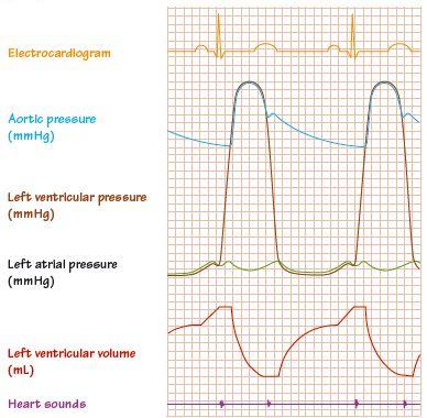
Fig. 3.5 The basic elements of the cardiac cycle.
Stay updated, free articles. Join our Telegram channel

Full access? Get Clinical Tree


