Arrhythmia Surgery and Fontan Conversion


Arrhythmia Surgery and Fontan Conversion


Constantine Mavroudis

Barbara J. Deal

Carl L. Backer


INTRODUCTION

Arrhythmia surgery in young patients is most commonly performed in the setting of concurrent repair of structural heart disease, and rarely performed in patients with structurally normal hearts with arrhythmias refractory to ablations and medications. As the success of surgical repairs of congenital heart disease has improved survival, the late sequelae of repaired congenital heart disease include late hemodynamic/structural problems, arrhythmias, and congestive heart failure. By adulthood, the development of atrial arrhythmias in the setting of congenital heart disease is associated with a 50% increase in mortality, a twofold increase in stroke or congestive heart failure, and a tripling of the need for cardiac interventions. The incidence of reoperations for complex lesions parallels the incidence of arrhythmias and underscores the complex electromechanical interactions in congenital heart disease. In patients with congenital heart disease, arrhythmia surgery may be performed as therapy for coexisting arrhythmias, or prophylactically to reduce the risk of developing late atrial arrhythmias. Efforts to minimize arrhythmia occurrence can be expected to reduce the morbidity related to arrhythmias and hospitalizations and may alter the risk of sudden death. It has been our practice to integrate anti-arrhythmia therapies into all primary repairs as needed, and to view any reoperation for congenital heart disease as an opportunity to improve both the electrical as well as the hemodynamic status of the patient. A unique subset that we will focus on in this chapter is the patient with a prior atriopulmonary Fontan. These patients undergo a Fontan conversion with arrhythmia surgery.

Arrhythmia surgery in patients with congenital heart disease is challenged by the range of anatomic variants, arrhythmia types, and intramyocardial scar location. Anatomic variants include unusual atrioventricular (AV) as well as ventricle to great vessel connections, juxtaposition of the atrial appendages, anomalous pulmonary and systemic venous return among a myriad of other complex lesions. Arrhythmia types can generally be grouped into specific mechanisms, but location and ablation can be perplexing and demanding as each of the arrhythmias is ablated by different procedures indicated by the arrhythmia substrate. Scar formation is generally induced by previous reparative operations that are required to achieve a separated pulmonary and systemic circulation, but not always. Scar formation leading to intractable arrhythmias may occur owing to intracavitary hemodynamic jet lesions, atrial dilatation, myocardial tumors, and cardiomyopathy.

The purpose of this chapter is to review arrhythmia mechanisms and operative techniques for arrhythmia surgery, with attention to particular challenges associated with congenital heart disease, including resternotomy techniques for safe mediastinal reentry. The historical review is based on our previous publications in this field involving 248 patients between 1987 and 2010 from Children’s Memorial Hospital in Chicago and the Cleveland Clinic Children’s Hospital (Tables 105.1, 105.2 and 105.3).


ARRHYTHMIA MECHANISMS AND ARRHYTHMIA SURGERY TECHNIQUES

A simplistic approach to the categorization of arrhythmias is summarized in Table 105.4. The most common types of arrhythmias associated with congenital heart lesions are atrial reentry tachycardia (ART), atrial fibrillation (AF), accessory connections, AV nodal reentry tachycardia (AVNRT), focal or automatic atrial tachycardia, and ventricular tachycardia (VT). Reentry is the most common mechanism for clinical arrhythmias and accounts for more than 60% of late postoperative arrhythmias. A reentrant rhythm requires a circuit, with unidirectional block and slowed conduction allowing the electrical impulse to “reenter” the previously blocked area. Reentry may occur in the atria (atrial flutter or atrial reentry), in the ventricles (postinfarction or scar-related VT), or involve all chambers (reciprocating tachycardia utilizing an accessory connection). Reentrant arrhythmias may be initiated or terminated by pacing, allowing the reproduction of the arrhythmia for elucidation of the mechanism in the catheterization laboratory or operating room. Most reentrant arrhythmias are suitable for catheter or surgical ablation, by interrupting a key component of the reentrant circuit. The majority of reentrant circuits are large, or “macro-reentrant,” such as atrial flutter. In typical atrial flutter, the electrical impulse courses up the atrial septum and down the right atrial free wall, with slowed conduction in the isthmus of tissue between the coronary sinus ostium and the tricuspid valve. In some instances, the reentrant circuit is more circumscribed, or “micro-reentrant,” and may be amenable to targeted focal ablative techniques in a discrete region. In contrast, automatic arrhythmias are local areas of increased firing and cannot be terminated by pacing or cardioversion; therapy is directed at slowing the rate of firing, or eliminating the specific focus of automaticity with ablation or resection. Examples of automatic rhythms include sinus rhythm, junctional ectopic tachycardia, and accelerated ventricular rhythms. The correct characterization of the arrhythmia circuit is essential to the success of arrhythmia surgery, as the most perfectly executed maze procedure for AF will not eliminate a focal source of atrial automaticity.

Surgical and transcatheter techniques have been highly effective in ablating these arrhythmias in pediatric and adult patients with congenital heart disease and normal anatomic hearts. Difficulty with transcatheter ablation techniques arises with complex anatomic variants, or
complexities posed by limitations of venous access or surgical baffles or conduits. Patients with double-outlet right ventricle with subaortic conus can harbor an accessory connection in the subaortic conus between the discontinuous aortic annulus and mitral annulus, a situation that does not exist in normal hearts where the ablative techniques have been standardized. Patients with heterotaxy syndrome with either a functionally univentricular heart or two separate ventricles may have absence of the coronary sinus, presence of a left superior vena cava, separate atrial entry of the hepatic veins, and/or juxtaposition of the atrial appendages, in addition to surgical baffles limiting access to regions of the atria. Patients with congenitally corrected transposition of the great arteries (ccTGA) have a propensity to display multiple accessory connections in association with Ebstein’s anomaly of the systemic tricuspid valve. In addition to these anatomic variants, repaired tetralogy patients have a high incidence of ART caused by atrial dilatation and scar formation, as well as VT in the area of the ventricular septal defect closure and right ventriculotomy. Transcatheter ablation in the older Fontan patient is hampered by both the multiplicity of arrhythmia mechanisms and circuits, location of some arrhythmia circuits in the left atrium or under surgical patches over the atrial isthmus, as well as the marked atrial hypertrophy limiting the ability to create transmural conduction block. The contemporary congenital heart surgeon should have a comprehensive understanding of all arrhythmia types and the potential methods of ablation, as well as hurdles of anatomic complexity for arrhythmia surgery if a cardiac operation or reoperation for an anatomic repair/re-repair is required in a patient with arrhythmias.


Table 105.1 Arrhythmia Surgery and Congenital Heart Disease Repair: Children’s Memorial Hospital and the Cleveland Clinic Children’s Hospital Experience 1987 to 2010 (n = 248)


Mean age


21.3 ± 10.9 y (range: 7 d to 64 y)


Associated congenital heart disease


236 patients


Resternotomy


83%


Functionally univentricular heart


67%


Two ventricles


33%


Symptomatic due to arrhythmia recurrences


100%


(Reproduced with permission from Mavroudis C, Deal BJ, Stewart RD. Operative techniques in association with arrhythmia surgery in patients with congenital heart disease. World J Pediatr Congen Heart Surg 2013;4:85-97.)


Table 105.2 Arrhythmia Types


N


Macro-reentrant atrial tachycardia


117


Right-sided


104


Right- and left-sided


13


Atrial fibrillation


86


AV nodal reentry tachycardia


6


Accessory connections (WPW)


19


Focal (automatic) atrial tachycardia


6


Ventricular tachycardia


14


AV, atrioventricular; WPW, Wolff-Parkinson-White syndrome. (Reproduced with permission from Mavroudis C, Deal BJ, Stewart RD. Operative techniques in association with arrhythmia surgery in patients with congenital heart disease. World J Pediatr Congen Heart Surg 2013;4:85-97.)


Table 105.3 Associated Diagnoses


N


Functionally univentricular heart


164


Tetralogy of Fallot/PA-VSD/DORV


22


Transposition of great arteries (1 ccTGA)


11


VSD (1 multiple)


7


AV canal (5 partial)


7


ASD (1 sinus venosus)


5


PA/PS-IVS


5


Aortic stenosis/atresia


3


Ebstein’s anomaly


4


Mitral regurgitation


3


Truncus, Uhl, absent PV, Scimitar


5


Structurally normal heart


12


ASD, atrial septal defect; AV, atrioventricular; ccTGA, congenitally corrected transposition of the great arteries; DORV, double-outlet right ventricle; IVS, interventricular septum; PA, pulmonary atresia; PS, pulmonary stenosis; PV, pulmonary valve; VSD, ventricular septal defect. (Reproduced with permission from Mavroudis C, Deal BJ, Stewart RD. Operative techniques in association with arrhythmia surgery in patients with congenital heart disease. World J Pediatr Congen Heart Surg 2013;4:85-97.)


ARRHYTHMIA-SPECIFIC SURGERY TECHNIQUES

The basic techniques for arrhythmia surgery include dissection, linear or focal ablation (sometimes in tandem with dissection), and maze procedures. Focal or micro-reentrant tachycardias require targeted dissection or ablation of the localized arrhythmogenic focus. Accessory connections are treated with discrete ablation, or endocardial dissection along the AV annulus for multiple or complex-branching connections, as seen with Ebstein’s anomaly of the tricuspid valve. maze procedures are designed to eliminate macro-reentrant
circuits in the atria, while preserving intact AV conduction and atrial transport function, and may be performed using a combination of resection and linear ablative lesions. The right atrial maze procedure is performed for atrial reentry or atrial flutter, and both a right and left atrial maze procedure is performed for AF. The right atrial maze procedure alone is not adequate to eliminate AF, which is primarily of left atrial origin.


Table 105.4 Types of Arrhythmias


Arrhythmia


Examples


Bradycardia


Sinus node dysfunction; atrioventricular block


Tachycardia


Supraventricular


Atrial flutter/reentry


Atrial fibrillation


Focal


Accessory connection (WPW)


AV nodal reentry or junctional


Ventricular


Secondary: inotropes, electrolytes, injury


Scar-related


Ion channelopathy mediated


Arrhythmic:


Bradycardic: asystole, or complete atrioventricular block


Atrial tachycardia


Sudden death


Ventricular tachycardia/fibrillation


Nonarrhythmic: pulmonary embolus, myocardial ischemia, electromechanical dissociation


Cerebrovascular accident


AV, atrioventricular; WPW, Wolff-Parkinson-White syndrome.


MACRO-REENTRANT ATRIAL TACHYCARDIA; ATRIAL REENTRY TACHYCARDIA

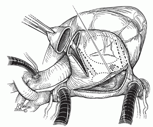
As noted previously, typical atrial flutter is a macro-reentrant circuit of the right atrium with an area of slowed conduction between the inferior vena cava (IVC), tricuspid valve, and the coronary sinus, characterized by saw-toothed flutter waves visible on electrocardiogram, and a relatively rapid atrial rate. In repaired congenital heart disease, macro-reentrant atrial tachycardia may occur in either atrium and may involve areas of slow conduction posed by electrical or surgical scar (crista terminalis, atrial septal defect patch) or anatomic orifices (inferior or superior vena cava, atriopulmonary anastomosis, entry of pulmonary veins or anomalies of venous return); in this setting the arrhythmia is labeled “atrial reentrant” or “intra-atrial reentrant” or “scar-mediated atrial reentrant” by various authors. Typical flutter waves are not usually apparent, and atrial rates are slower than typical atrial flutter, resulting in discrete P waves visible on electrocardiogram with variable AV conduction. Ablative lesions are used to interrupt the macro-reentrant circuit in the areas of slowed conduction with anatomic barriers, most commonly at the inferior right atrial isthmus, defined as the area between the coronary sinus, the tricuspid annulus, and the IVC. The ablative lesion, therefore, transforms an area of slow conduction to an area of no conduction, thereby eliminating the circuit. In normal hearts, the therapeutic lesions are noted in Figure 105.1.


Fig. 105.1. Right-sided maze procedure used in patients with atrial reentry tachycardia and a two-ventricle circulation. Some of the lesions are “cut and sew,” others are performed with a cryoablation catheter (-160°C × 1 minute). (Reproduced with permission from Mavroudis C, Deal BJ, Backer CL. Arrhythmia surgery in association with complex congenital heart repairs excluding patients with Fontan conversion. Semin Thorac Cardiovasc Surg Pediatr Card Surg Annu 2003;6:33-50.)

Multiple areas of slow conduction may exist in patients with congenital heart disease from prior incisions, patches, or wall stress and the ablative lesions target the bridges between anatomic barriers. Identification of the relevant macro-reentrant circuits and targeting the potential areas of slowed conduction suitable for linear ablations (connecting anatomic barriers) are key to successful ablation. While the anatomic variants are many, the tenets of ablative therapy are constant, namely, that lines of block are established between two or more anatomic barriers. For right ART in the setting of congenital heart disease, the surgeon is concerned with the inferior and superior vena cavae, hepatic venous entry,
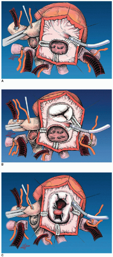
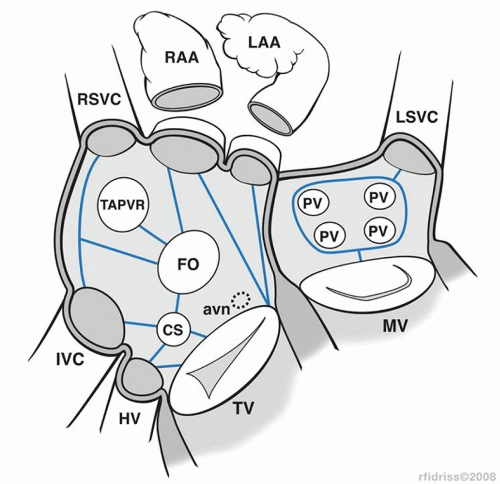
AV valve, coronary sinus, atrial appendage, and the fossa ovalis or atrial septal patch. When anatomic barriers are absent or anomalous, creative application become necessary by delivering ablative lesions using the guidelines mentioned previously. For example, since there is no tricuspid valve in tricuspid atresia, cryoablation lesions are applied as noted in Figure 105.2A. Figure 105.2B and 105.2C demonstrates the lesions that are required for single right ventricle/mitral atresia and functionally univentricular hearts with unbalanced AV canal, respectively. Figure 105.3 is a complex anatomic diagrammatic representation of the potential anomalies of systemic and pulmonary venous return, juxtaposed atrial appendages and proposed lines of block that are recommended for right- and left-sided maze procedures. The ablative lesions noted in Figure 105.3 are designed to be guidelines that are executed based on the preoperative electrophysiologic study and the type of arrhythmia that has been characterized. As an example, biatrial maze operations are performed if suture lines extend into the left atrium or if there is anomalous left superior vena caval drainage.


Fig. 105.2. These are a series of illustrations of the use of cryoablation in patients undergoing Fontan conversion. (A) The modified right-sided maze procedure in a patient with tricuspid atresia. (B) The modified right-sided maze procedure in a patient with double-outlet right ventricle and mitral atresia. (C) The modified right atrial maze procedure in a patient with a functionally univentricular heart (unbalanced atrioventricular septal defect). (Reproduced with permission from Mavroudis C, Backer CL, Deal BJ, Johnsrude C, Strasburger J. Total cavopulmonary conversion and maze procedure for patients with failure of the Fontan operation. J Thorac Cardiovasc Surg 2001;122:863-871.)


FOCAL OR AUTOMATIC ATRIAL TACHYCARDIA

Focal (automatic) atrial tachycardia is characterized by a localized area of electrical impulse generation, which may be caused by discrete micro-reentry or by an automatic focus. Either form of focal tachycardia inhibits the slower rate of normal sinus node firing and results in tachycardia. The electrical impulse from this ectopic focus is conducted and propagated through the atria, often with prolongation of the PR interval, resulting in stimulation of the AV node and ventricles. Surgical treatment of focal (automatic) atrial tachycardia usually requires resection and/or ablation of atrial tissue; more extensive
areas of focal tachycardia may require electrical anatomic isolation. Favorable outcomes have been achieved with cryoablation and excision of automatic foci. Automatic right atrial foci are often localized to the right atrial appendage and can be resected. Multiple ectopic foci resulting in arrhythmia recurrence stimulated surgeons to use more extensive and creative techniques such as pulmonary vein isolation, left atrial isolation, right atrial isolation, and His bundle ablation with pacemaker insertion in difficult cases.


Fig. 105.3. Schematic representation of the possible lines of ablation to treat macro-reentrant atrial tachycardia in the presence of various atrial anomalies associated with complex congenital heart disease. avn, atrioventricular node; CS, coronary sinus; FO, foramen ovale; HV, hepatic vein; IVC, inferior vena cava; LAA, left atrial appendage; LSVC, left superior vena cava; MV, mitral valve; PV, pulmonary valve; RAA, right atrial appendage; RSVC, right superior vena cava; TAPVR, total anomalous pulmonary venous return; TV, tricuspid valve. (Reproduced with permission from Mavroudis C, Deal BJ, Backer CL, Tsao S. Arrhythmia surgery in patients with and without congenital heart disease. Ann Thorac Surg 2008;86:857-868.)

We have successfully performed atrial appendage resection for automatic atrial tachycardia in two neonates and five older children (Fig. 105.4). One neonate underwent a concomitant Norwood procedure for hypoplastic left heart syndrome and the other, with a normal heart, was spared extracorporeal membrane oxygenation (ECMO) after the arrhythmia site (right atrial appendage) was resected and ablated in the operating room. Of the five older children, three underwent concomitant atrial (n = 2) and ventricular (n

Only gold members can continue reading. Log In or Register to continue

Stay updated, free articles. Join our Telegram channel

Jun 15, 2016 | Posted by in CARDIAC SURGERY | Comments Off on Arrhythmia Surgery and Fontan Conversion

Full access? Get Clinical Tree

Get Clinical Tree app for offline access