Anatomy of the Thorax

CHAPTER 1 Anatomy of the Thorax


The thorax is the upper part of the trunk, bounded by the diaphragm inferiorly, the thoracic inlet superiorly, and the thoracic cage between. Vital organs, such as the heart and lungs, reside completely within the thoracic cavity, and other, equally vital organs, such as the aorta and esophagus, extend into the abdominal cavity. The thorax can be divided into a right and left hemithorax, separated by the mediastinum. This chapter gives an overview of the anatomy of the thoracic cavity and its contents.


THORACIC CAGE


The cylindrical thoracic cage has two main purposes: it provides protection for the underlying organs, and the dynamic interactions of the bony and muscular components of the chest wall allow changes in respiratory volumes.


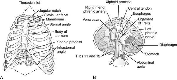
The bony thorax has two apertures, or openings: the superior thoracic aperture, often referred to as the thoracic inlet or cervicothoracic junction, and the inferior thoracic aperture (Fig. 1-1A).


Cervicoaxillary Canal


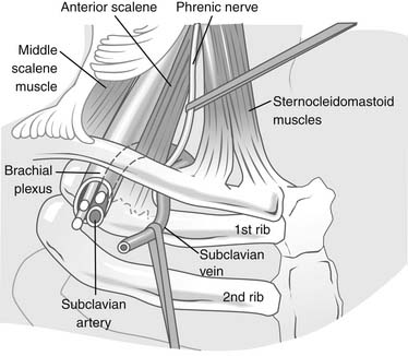
The cervicoaxillary canal is bounded by the first rib inferiorly, the clavicle superiorly, and the costoclavicular ligament medially (Fig. 1-2). The structures that pass through this space include the subclavian vein and artery and the brachial plexus.


The subclavian artery passes over the first rib and goes between the scalenus anterior and scalenus middle muscles. Before passing over the first rib on its way out the cervicoaxillary canal, it gives off branches, including the thyrocervical, internal mammary, and vertebral arteries. It becomes the axillary artery after it exits from behind the pectoralis minor muscle. Compression of this artery may lead to poststenotic dilation, stenosis, aneurysmal formation, or occlusion.


The subclavian vein travels from the arm and goes behind the pectoralis minor muscle before going between the first rib and clavicle. This space is bounded medially by the costoclavicular ligament and laterally by the scalenus anterior muscles as it inserts onto the first rib. Compression of the vein occurs in this area and can lead to stenosis and occlusion.


The brachial plexus is composed of nerve roots from the C5 through the T1 vertebral foramina. These nerve roots join to form the superior trunk (C5 and C6), the middle trunk (C7), and the inferior trunk (C8 and T1). These trunks separate into anterior and posterior divisions that fuse again to form lateral, medial, and posterior cords of the brachial plexus. The plexus travels through the tunnel formed by the anterior and middle scalene muscles. It travels between the first rib and the clavicle and finally under the pectoralis minor muscle to gain access to the arm.


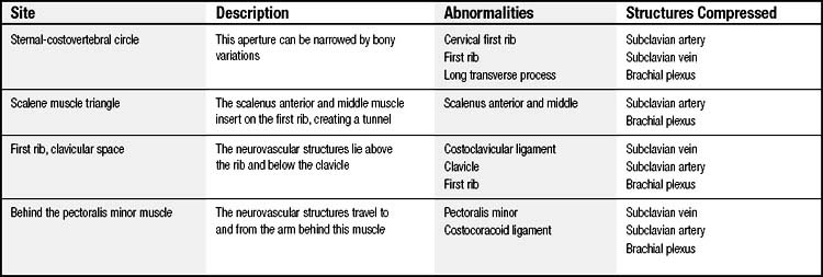
In the normal anatomy, there is ample room for each of these structures. There are four major areas in which the vessels or nerves can be compressed (Table 1-1).This topic will be discussed in greater detail in Chapter 26.


Diaphragm


The diaphragm, at the inferior thoracic aperture, separates the thoracic cavity from the abdominal cavity. The inferior thoracic aperture, which slopes inferoposteriorly, is limited by the 12th thoracic vertebra posteriorly, the 12th pair of ribs and costal margins anterolaterally, and the xiphisternal joint anteriorly.


The diaphragm is a curved musculotendinous sheet that is mainly convex toward the thoracic cavity. It is a continuous sheet of muscle with low posterior and lateral attachments and high anterior attachments. The central tendon is a thin but strong aponeurosis. The domes of the diaphragm descend 2 cm during quiet respiration and may travel as much as 10 cm during heightened respiratory requirements. During expiration, the right diaphragm may rise as high as the level of the nipple, whereas the left rises to a level one rib space lower. With maximal inspiration, the diaphragm flattens against the abdominal contents. The right diaphragm may flatten to the level of the 11th rib, whereas the left may flatten to the level of the 12th rib. The right diaphragm has to work against the liver, whereas the left needs to push only against the stomach and spleen. Therefore, the right diaphragm has significantly stronger fibers than the left.


The costal diaphragm receives its blood supply from the lower five intercostal and subcostal arteries, and the central portion is supplied by the phrenic arteries. The phrenic arteries may arise directly from the aorta, above the celiac axis, as a common trunk or individually. Occasionally, they arise from the celiac or renal arteries. The sole motor supply to the diaphragm is the phrenic nerve. It also supplies sensory fibers to the parietal and peritoneal pleura overlying the diaphragm. This accounts for the diaphragmatic irritation that is sometimes interpreted as ipsilateral shoulder pain.


The diaphragm has several openings through which structures can transverse from one cavity to another (see Fig. 1-1B). The inferior vena cava hiatus lies anteriorly and to the right of the midline, at the level of vertebra T8. The right phrenic nerve travels through this hiatus, and the left phrenic nerve penetrates laterally through its own opening at the same level. The esophageal aperture is at the level of the T10 vertebra, and the right and left vagal trunks that adhere to it enter the abdomen along with the esophagus.


The aortic aperture, at the T12 vertebral level, is formed by the interdigitating fibers (median arcuate ligament) of the right and left diaphragmatic crura. The azygos and hemiazygos veins and the thoracic duct also pass through this opening.


The greater and lesser splanchnic nerves gain access to the abdominal cavity via two small apertures in each of the crura, and musculophrenic branches of the internal mammary artery penetrate the diaphragm through small apertures near its connection with the costal cartilages of ribs seven to nine.


Diaphragmatic hernias can develop through known openings, such as a hiatal hernia through the esophageal hiatus, or through congenital defects. The most common congenital abnormality is a Bochdalek hernia, in which abdominal contents go through a posterolateral defect. This is seen more commonly on the left. A Morgagni hernia, on the other hand, occurs with a defect in the anterior aspect of the diaphragm just lateral to the xiphoid.


The Bony Thoracic Cage


Sternum


The sternum or breastbone is made of cancellous bone and filled with hemopoietic marrow throughout life. The main parts, the manubrium and body, are connected by a secondary cartilaginous joint that normally never ossifies and contributes to movement of the ribs.


Up to puberty, the six segments, or sternebrae, are held together by hyaline cartilage. The central four fuse to form the body of the sternum between 14 and 21 years of age. The manubrium sterni (superiorly) and the xiphoid process (inferiorly) remain separate.


The manubrium receives the sternal ends of the clavicles in a shallow concave facet. The widest portion of the manubrium has bilateral costal incisurae that articulate with the first costal cartilage to form a primary cartilaginous joint. The second costal cartilage articulates with both the lower lateral ends of the manubrium and the body of the sternum, forming separate synovial joints. The muscular attachments of the manubrium include the sternocleidomastoid muscle, the sternohyoid and the sternothyroid superiorly, and the pectoralis major muscle anterolaterally. Most of the posterior surface is bare bone, which may be in contact with the brachiocephalic vein unless thymic remnants lie between.


The gladiolus, or body of the sternum, is slanted at a steeper angle than the manubrium; its articulation with that bone forms the sternal angle. Ossification of this joint, synchondrosis, in adult life may limit normal movement at this joint. The articular facets for ribs two to seven lie along the lateral border of the body of the sternum. These make single synovial joints with the costal cartilages. The facets for the sixth and seventh costal cartilages may coalesce, especially in females. The lateral border gives attachment to the anterior intercostal membrane and the internal intercostal muscles, and the pectoralis major arises anteriorly. Weak sternopericardial ligaments pass into the fibrous pericardium.


The cartilaginous xiphoid may be bifid or perforated, is of variable length, and usually ossifies in the 4th decade. The costoxiphoid ligaments prevent its displacement during diaphragmatic contractions.


Ribs


The 12 pairs of ribs are divided into the upper seven, which that are called true ribs or vertebrosternal ribs because they form complete loops between the vertebrae and the sternum, and the lower five ribs, which fail to reach the sternum and are considered false ribs. The eighth, ninth, and 10th ribs are called vertebrocostal because each of their costal cartilages articulates with the adjacent rib cartilage. Ribs 11 and 12 are free-floating or vertebral ribs because their only articulation is with their vertebrae.


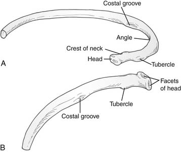
Ribs three to nine are classified as typical ribs and have a head, neck, and a shaft. The head has an upper and a lower articular facet divided by a crest for articulation with two adjacent vertebrae in synovial costovertebral joints, the lower facet articulating with the upper border of its own vertebra. The neck is flattened, with the upper border curling up into a prominent ridge—the crest. A tubercle projects posteriorly from the end of the neck and marks the junction of the neck and the body (Fig. 1-3).


The medial facet on the tubercle is covered with hyaline cartilage and makes a synovial joint with the transverse process of its own vertebra. The lateral facet receives the costotransverse ligament from the tip of its own transverse process. The shaft slopes downward and laterally for about 5 to 8 cm at an angle and then curves forward. Lateral to the angle, the lower border projects down as a sharp ridge, sheltering a costal groove. The angles of the ribs also correspond to the lateral extent of the erector spinae muscles of the back. The upper six ribs are bent into a tight curve so that the shaft has turned parallel with the neck of the rib. The lower six ribs have an opening out of the curve, which is completed by the long costal cartilages at the front of the chest. The fused cartilages of ribs seven to 10 course diagonally upward to the lower end of the sternum to form the subcostal angle. The anterior ends of the ribs have a concave fossa that is plugged by the costal cartilage in an immovable primary cartilaginous joint.


The first rib is exceptionally broad and short and very highly curved. The head is small and has a single facet for the synovial joint it makes with the upper part of the body of the T1 vertebra. The prominent tubercle is a fusion of the tubercle and the angle, and its medial facet forms a synovial joint with the first transverse process. The lateral prominent part of the tubercle receives the lateral costotransverse ligament and the costalis and longissimus parts of the erector spinae. Near the middle of the shaft, the sulci for the subclavian vessels are identified by the scalene tubercle, a spur that attaches the scalenus anterior muscle.


MUSCLES OF THE THORAX


The muscles of the chest wall serve to protect the contents of the thoracic cavity, and they assist the movements of the thorax and upper extremities (Fig. 1-4). The 17 muscles of the chest wall are not discussed in detail here, but Table 1-2 lists them with their innervations and sites of attachments. Here we focus on muscle groups that are used in chest wall reconstruction. The latissimus dorsi, pectoralis major, serratus anterior, trapezius, rectus abdominis, and external oblique are the six major muscles that are available for reconstruction (Table 1-3).


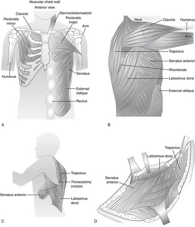
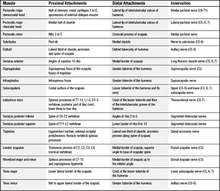
Table 1–3 Arterial Supply of Muscles Used in Chest Wall Reconstruction


Muscle Arterial Supply
Latissimus dorsi Thoracodorsal artery
Pectoralis major Thoracoacromial, lateral thoracic perforators, internal mammary artery, lateral intercostals artery
Serratus anterior Thoracodorsal artery, long thoracic artery
Trapezius Transverse cervical artery
External oblique Lower thoracic intercostal arteries
Rectus abdominis Superior and inferior epigastric arteries

The latissimus dorsi is the largest muscle of the thorax. It originates from the lower six thoracic spinous processes and from the lumbodorsal fiber, which is attached to the lumbar and sacral vertebrae. It also has fibers originating from the iliac crest. The muscle narrows and then inserts on the intertubercle groove of the humerus. It is supplied by the thoracodorsal artery, a branch of the subscapular artery. The subscapular artery arises from the axillary artery and gives a branch to the serratus anterior muscle before supplying the latissimus dorsi. The artery can be found on the undersurface of the latissimus dorsi. The mobile arterial supply and excellent musculocutaneous collaterals allow this muscle to be moved with or without its overlying skin.


The pectoralis major muscle arises from the sternum, clavicle, and first seven ribs. It inserts on the bicipital humeral groove. It receives arterial supply from a pectoral branch of the thoracoacromial artery arising midclavicularly. It also receives some blood supply from the internal mammary, lateral intercostals, and lateral thoracic perforators. This flap is most frequently used for sternal defects.


Located between the latissimus dorsi and pectoralis major muscles is the serratus anterior muscle. This small muscle originates from the superior borders of the eighth through tenth ribs. It inserts on the tip of the scapula. Its blood supply is from a branch of the thoracodorsal artery and from the long thoracic artery. It can be used for intrathoracic purposes such as bronchial stump coverage or muscle interposition between the trachea and esophagus after a tracheo-esophageal fistula repair.


The trapezius muscle arises from the occipital bone and from the spinous process of the seventh cervical vertebra and all the thoracic vertebrae. It inserts on the lateral aspect of the clavicle, the acromion process, and the spine of the scapula. It receives its blood supply from the transverse cervical artery. Although it is a large muscle, the use of the trapezius is limited to reconstruction of the upper thoracic cavity.


Arising from the inferior borders of the fourth through twelth ribs and inserting on the iliac crest is the external oblique muscle. It accepts blood supply from the lower thoracic intercostal arteries. Unlike the trapezius muscle, its uses are limited to the thoracic cavity below the inframammary crease.


The rectus abdominis muscle spans the entire anterior abdominal wall. It originates at the pubic crest and rises superiorly to insert on the fifth through seventh rib cartilages and xiphoid process. The superior and inferior epigastric arteries supply this vast muscle. It has been used for reconstruction of the anterior thoracic cavity, most notably after breast surgery.


Intercostal Muscles


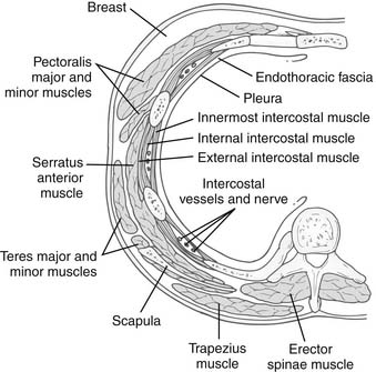
Fibers of the intercostalis externi muscle arise from the sharp lower border of the rib above and course inferomedially (i.e., in the direction of the fingers when the hands are put into the front pockets of trousers) to the smooth upper border of the rib below. Anteriorly, it is replaced by the anterior intercostal membrane. Between the bony ribs is muscle; between the costal cartilages is membrane. In the lower spaces, the muscle interdigitates with the fibers of the external oblique (Fig. 1-5).


The intercostalis interni muscle runs from the lower costal groove of ribs one to 11, to the upper surfaces of ribs two to 12 downward and backward. Anteriorly, they extend up to the sternum but posteriorly only up to the angles of the rib. Beyond that, they are replaced by the internal intercostal membrane, which attaches to the tubercle of each rib and vertebra.


The innermost or transversus layer is divided into three groups: the innermost intercostals (anterolateral), subcostal (posterior), and transversus thoracis (anteromedial) muscles. The fibers run downward and backward, as in the internal intercostal muscles. The subcostal muscles lie in the paravertebral gutter, are better developed inferiorly, and cross more than one space. The transversus thoracis was formerly called the sternocostalis, a more appropriate name. Digitations arise from the sternum bilaterally to the costal cartilages of ribs two to six.


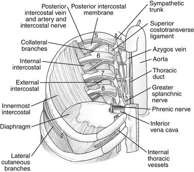
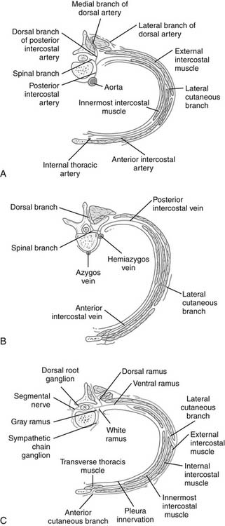
The intercostalis intimi muscles also traverse more than one space and are better developed in the lower lateral spaces. In the plane between the innermost and outer two layers runs the neurovascular bundle. From above downward, the order is vein, artery, and nerve (mnemonic: VAN). Beyond the angle of the rib posteriorly, they are protected by the downward projection of the lower border of the rib. Hence, for a thoracotomy, the periosteum is stripped off the upper half of the rib, avoiding the lower border and the neurovascular bundle (Fig. 1-6).


As in the rest of the body, the mixed spinal nerve is formed from a dorsal and a ventral root, the dorsal root being sensory and the ventral root containing somatic motor neurons. As it emerges from the intervertebral foramina, it branches into a dorsal and a ventral ramus. The dorsal ramus of the thoracic spinal nerve supplies the paravertebral back muscles and skin of the back. The ventral ramus communicates with the sympathetic chains via white rami communicantes (postganglionic fibers). Beyond this point, the true intercostal nerve lies just superficial to the parietal pleura in the endothoracic fascia. It gains the costal groove between the innermost intercostal and the internal intercostal muscles near the angle of the rib, where a collateral branch is given off. This small branch supplies the muscles of the space, the parietal pleura, and the periosteum of the rib. The main nerve itself has muscular branches, a lateral cutaneous branch, and a terminal anterior cutaneous branch. The lateral cutaneous branch given off along the midaxillary line gives off anterior and posterior branches to supply the skin over that space. Just lateral to the sternal margin, the terminal anterior cutaneous branches of the upper six nerves pierce the internal intercostal muscles, the external intercostal membrane, and the pectoralis major to reach skin. The lower five intercostal nerves slope downward behind the costal margin into the neurovascular plane of the abdominal wall. The subcostal or 12th thoracic nerve leaves the thorax by passing behind the lateral arcuate ligament and the subcostal artery and vein (Fig. 1-7). The cutaneous branches of each dermatome tend to overlap considerably; hence anesthesia after thoracic incisions is quite rare unless multiple intercostal nerves have been damaged. The first intercostal nerve is very small and supplies no skin, lacking both lateral and anterior cutaneous branches.


Two sets of intercostal arteries, the posterior and the anterior, are responsible for supplying the intercostal spaces. Their course and branching pattern closely conform to those of the intercostal nerves. The posterior intercostal arteries are branches of the descending thoracic aorta except in the first two spaces, where they are branches of the supreme intercostal artery, given off by the costocervical trunk of the second part of the subclavian artery. The aortic branches lie on the left side of the mediastinum; consequently, the right posterior intercostal arteries are longer, can be easily dissected in the left chest, and are seen best while operating on descending thoracic aortic aneurysms.


The roots of all 12 pairs of posterior intercostal arteries give rise to a dorsal branch that supplies the vertebrae, spinal cord, and deep muscles of the back. Like the nerve, the intercostal artery gives off a collateral branch, the largest muscular branch that runs along the upper border of the rib below the space. After gaining the costal groove, near the midaxillary line, it gives off the lateral cutaneous branch. The subcostal artery or the 12th thoracic posterior intercostal artery follows a similar course but has no collateral branch.


The anterior intercostal arteries are branches of the internal thoracic arteries from the first part of the subclavian artery. The internal thoracic artery runs anterior to the transversus thoracis and on the internal surface of the costal cartilages and the internal intercostal muscles. About a finger’s breadth from the border of the sternum running vertically downward, it gives off two anterior intercostal arteries in each space. At the costal margin, below the sixth costal cartilage, it divides into the superior epigastric and musculophrenic arteries. The anterior intercostal arteries are smaller than the posterior intercostal arteries with which they anastomose and run predominantly along the lower border of each costal cartilage in the same fascial neurovascular plane. In the lower spaces, they are branches of the musculophrenic artery. There are no true anterior intercostal arteries in the last two spaces.


The internal thoracic arteries also give branches to the mediastinum, the thymus, the pericardium, and the sternum, and especially large perforating branches in the second to fourth space, the predominant supply to the lactating breast in females.


In each space there are one posterior and two anterior intercostal veins, designated by names identical to the arteries that they accompany. They lie above the artery and the nerve throughout their course (VAN) in the intercostal space. The anterior veins drain into the musculophrenic and internal thoracic veins. The vein of the first space or the supreme intercostal vein, posteriorly, may be a tributary of the brachiocephalic, vertebral, or superior intercostal vein. The superior intercostal vein is formed by the posterior intercostal veins of the second, third, and sometimes the fourth spaces. This drains into the azygos vein on the right side, and on the left side it arches over the aorta, superficial to the vagus and deep to the phrenic to open into the left brachiocephalic veins. Subcostal veins join the ascending lumbar veins and ascend on the left side as the hemiazygos and on the right side as the azygos vein draining the lower eight spaces. The blood in the hemiazygos vein drains across the midline, through median anastomoses into the azygos vein.


The internal thoracic vessels are part of the anastomotic chain that links the subclavian artery and brachiocephalic veins to the external iliac vessels. The intercostal vessels in turn connect to the descending aorta and azygos system of veins. In the presence of obstruction to flow, these anastomoses provide alternative channels for arterial and venous blood flow.


< div class='tao-gold-member'>

Stay updated, free articles. Join our Telegram channel

Jul 30, 2016 | Posted by in CARDIAC SURGERY | Comments Off on Anatomy of the Thorax

Full access? Get Clinical Tree

Get Clinical Tree app for offline access