Case 4
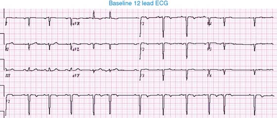
Baseline 12-Lead ECG
The baseline ECG demonstrates an abnormal P-wave axis, premature supraventricular complexes, and right-axis deviation of the QRS complexes. There is abnormal R wave progression in the precordial leads. The constellation of findings is suggestive of dextrocardia.
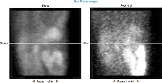
Raw Planar Images
The images were acquired over 180 degrees from the 45-degree right posterior oblique to 45-degree left anterior oblique orientations. Note that the heart is located in the right side of the chest, consistent with dextrocardia. The liver is located on the left, consistent with situs inversus.


