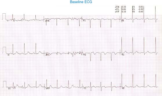
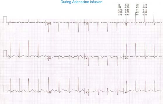
Case 18 A 54-year-old woman with inadequately controlled hypertension for the last several years complains of exertional angina at the outpatient office and is referred for pharmacologic stress/rest perfusion imaging. She is on oral atenolol 50 mg per day. She underwent 2-day rest/stress 99mTc-sestamibi perfusion imaging using 5-minute adenosine infusion as an outpatient study. Her heart rate increased from 83 to 93 beats/min and blood pressure from 185/99 to 179/98 mm Hg. She developed anginal chest pain during adenosine infusion. Her baseline and peak stress ECGs are shown. What is your interpretation? (Fig. 1a) (Fig. 1b) There is normal sinus rhythm with Q waves in aVL and V2, with T-wave inversion in leads aVL and V2-3. There is left ventricular hypertrophy. There is no ST-segment depression with adenosine infusion. (Video 1a) Only gold members can continue reading. Log In or Register to continue Share this: Share on X (Opens in new window) X Share on Facebook (Opens in new window) Facebook Related Related posts: Cardiac Neurotransmission Imaging: Single-Photon Emission Computed Tomography Myocardial Perfusion Imaging with Contrast Echocardiography Digital/Fast SPECT: Systems and Software Myocardial Perfusion: Magnetic Resonance Imaging Stay updated, free articles. Join our Telegram channel Join Tags: Clinical Nuclear Cardiology State of the Art and Future Direction Jun 11, 2016 | Posted by admin in CARDIOLOGY | Comments Off on 18 Full access? Get Clinical Tree
Case 18 A 54-year-old woman with inadequately controlled hypertension for the last several years complains of exertional angina at the outpatient office and is referred for pharmacologic stress/rest perfusion imaging. She is on oral atenolol 50 mg per day. She underwent 2-day rest/stress 99mTc-sestamibi perfusion imaging using 5-minute adenosine infusion as an outpatient study. Her heart rate increased from 83 to 93 beats/min and blood pressure from 185/99 to 179/98 mm Hg. She developed anginal chest pain during adenosine infusion. Her baseline and peak stress ECGs are shown. What is your interpretation? (Fig. 1a) (Fig. 1b) There is normal sinus rhythm with Q waves in aVL and V2, with T-wave inversion in leads aVL and V2-3. There is left ventricular hypertrophy. There is no ST-segment depression with adenosine infusion. (Video 1a) Only gold members can continue reading. Log In or Register to continue Share this: Share on X (Opens in new window) X Share on Facebook (Opens in new window) Facebook Related Related posts: Cardiac Neurotransmission Imaging: Single-Photon Emission Computed Tomography Myocardial Perfusion Imaging with Contrast Echocardiography Digital/Fast SPECT: Systems and Software Myocardial Perfusion: Magnetic Resonance Imaging Stay updated, free articles. Join our Telegram channel Join