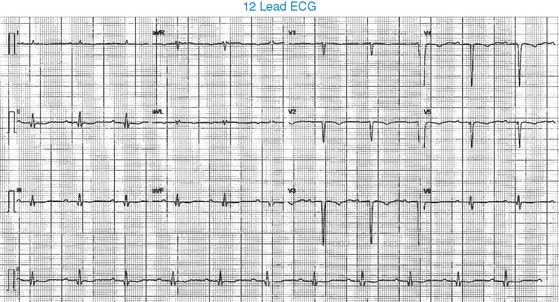
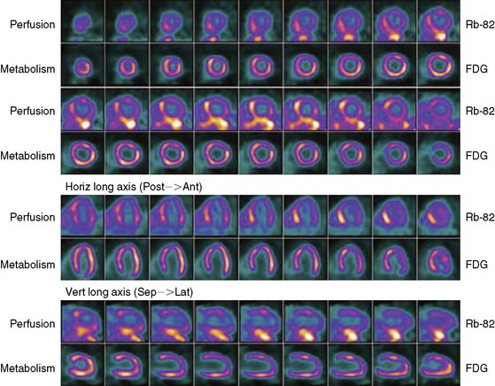
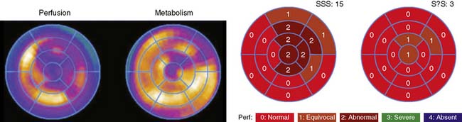
Case 15 A 66-year-old man with known severe three-vessel coronary artery disease (CAD) and ischemic cardiomyopathy was referred for myocardial viability assessment. Heart catheterization demonstrated severe left ventricular systolic dysfunction (left ventricular ejection fraction [LVEF] = 15% to 20%) and severe three-vessel CAD (75% proximal left anterior descending [LAD], 80% proximal circumflex, 80% mid–first obtuse marginal [OM1], 100% proximal right coronary artery [RCA]). 12-Lead ECG The baseline ECG demonstrates normal sinus rhythm, first-degree AV block, and Q waves suggestive of extensive anterior (and possibly inferior) infarction. Perfusion and Glucose Metabolism Rubidium-82 (82Rb) PET MPI demonstrates moderately severe and extensive perfusion defects in the anterior, anteroapical, and anterolateral regions. [18F]Fluorodeoxyglucose (18FDG) PET images demonstrate a mild localized area of reduced glucose metabolism in the anteroapical region. The “perfusion-metabolism mismatch” is consistent with preserved myocardial viability and predicts improvement of left ventricular systolic function with revascularization. Semiquantitative Assessment of Myocardial Perfusion and Glucose Metabolism The polar plots and 17-segment scoring results confirm that the regional reduction in myocardial perfusion (82Rb) is more severe and extensive than the reduction in glucose metabolism (18FDG). Only gold members can continue reading. Log In or Register to continue Share this: Share on X (Opens in new window) X Share on Facebook (Opens in new window) Facebook Related Related posts: Cardiac Neurotransmission Imaging: Single-Photon Emission Computed Tomography 18 Digital/Fast SPECT: Systems and Software Myocardial Perfusion: Magnetic Resonance Imaging Stay updated, free articles. Join our Telegram channel Join Tags: Clinical Nuclear Cardiology State of the Art and Future Direction Jun 11, 2016 | Posted by admin in CARDIOLOGY | Comments Off on 15 Full access? Get Clinical Tree
Case 15 A 66-year-old man with known severe three-vessel coronary artery disease (CAD) and ischemic cardiomyopathy was referred for myocardial viability assessment. Heart catheterization demonstrated severe left ventricular systolic dysfunction (left ventricular ejection fraction [LVEF] = 15% to 20%) and severe three-vessel CAD (75% proximal left anterior descending [LAD], 80% proximal circumflex, 80% mid–first obtuse marginal [OM1], 100% proximal right coronary artery [RCA]). 12-Lead ECG The baseline ECG demonstrates normal sinus rhythm, first-degree AV block, and Q waves suggestive of extensive anterior (and possibly inferior) infarction. Perfusion and Glucose Metabolism Rubidium-82 (82Rb) PET MPI demonstrates moderately severe and extensive perfusion defects in the anterior, anteroapical, and anterolateral regions. [18F]Fluorodeoxyglucose (18FDG) PET images demonstrate a mild localized area of reduced glucose metabolism in the anteroapical region. The “perfusion-metabolism mismatch” is consistent with preserved myocardial viability and predicts improvement of left ventricular systolic function with revascularization. Semiquantitative Assessment of Myocardial Perfusion and Glucose Metabolism The polar plots and 17-segment scoring results confirm that the regional reduction in myocardial perfusion (82Rb) is more severe and extensive than the reduction in glucose metabolism (18FDG). Only gold members can continue reading. Log In or Register to continue Share this: Share on X (Opens in new window) X Share on Facebook (Opens in new window) Facebook Related Related posts: Cardiac Neurotransmission Imaging: Single-Photon Emission Computed Tomography 18 Digital/Fast SPECT: Systems and Software Myocardial Perfusion: Magnetic Resonance Imaging Stay updated, free articles. Join our Telegram channel Join Tags: Clinical Nuclear Cardiology State of the Art and Future Direction Jun 11, 2016 | Posted by admin in CARDIOLOGY | Comments Off on 15 Full access? Get Clinical Tree