Acute kidney injury (AKI) remains a major concern in the field of TAVR and is associated with increased mortality, up to twofold to eightfold. Although the exact mechanism of post-TAVR AKI is unknown and seems to be multifactorial, contrast has been reported as a contributing factor. During TAVR, contrast is used for conventional aortic root angiography, for positioning of the TAVR valve device and assessing the peripheral vasculature. To our knowledge, for the first time, we report transfemoral TAVR performed with zero contrast using fluoroscopy and ultrasound guidance. This was done in two patients with symptomatic, severe aortic stenosis, and significant chronic kidney disease (stage III) who were deemed high risk for surgical aortic valve replacement after discussion in the multidisciplinary Heart Team meeting.
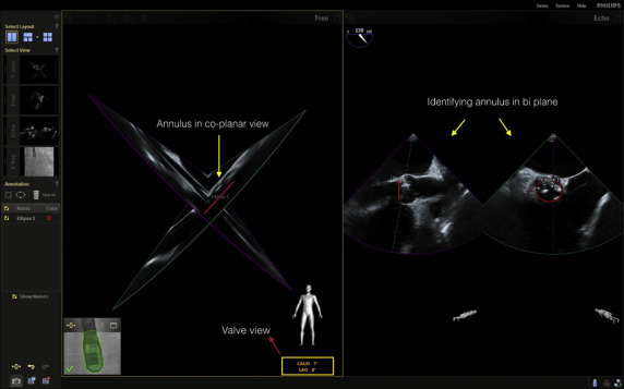
EchoNavigator system (Phillips Healthcare, Best, The Netherlands) is a novel imaging technology that allows fusion of real-time 2-dimensional (2D) and 3D echocardiography images on the fluoroscopy screen. It has the ability to identify the optimal perpendicular valve view without use of contrast ( Figure 1 ), can provide a road map to assist crossing of the aortic valve in difficult cases, and enables overlay of echocardiographic images on the x-ray screen so that the soft tissue characterization of echocardiography is combined with excellent device visualization of x-ray to assure precise device deployment. Determining the optimal valve view is a crucial prerequisite for accurate positioning of the bioprosthetic valve to avoid potentially life-threatening complications. Although aortic root angiography is conventionally used to identify the valve view, EchoNavigator can be solely used for this purpose. Moreover, to further minimize exposure to contrast, 3D transesophageal echocardiography, which is the standard technique in our center, was used for valve sizing.


Stay updated, free articles. Join our Telegram channel

Full access? Get Clinical Tree


