Zenker’s Diverticulum with Previous Left Carotid Endarterectomy
Presentation
A 72-year-old man presents with difficulty swallowing and regurgitation of previously swallowed solid foods.
His past medical history is significant for hypertension, coronary artery disease, and stroke. His past surgical history includes a left carotid endarterectomy 3 years ago. Review of systems is significant for weight loss of 15 pounds during the past year. A year ago, the patient suffered a stroke that resulted in right hemiparesis, which has improved with rehabilitation. The patient has had persistent dysphagia and reports feeling that the food is getting stuck in his throat. In addition, he frequently regurgitates undigested food, has foul-smelling breath, and was recently admitted to the hospital for pneumonia.
On physical examination, the patient appears emaciated and debilitated. He is afebrile and hemodynamically stable. There is no cervical adenopathy, and the thyroid gland is not enlarged. A left cervical scar is noted from previous carotid surgery. The lungs are clear, and his cardiac tones are normal. He has moderate right hemiparesis from his stroke. The rest of the examination is unremarkable.
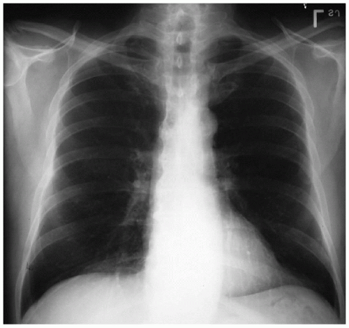
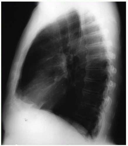
▪ Chest X-rays
Chest X-ray Report
Chest x-rays demonstrate clear lung fields. No masses are evident. The trachea is slightly deviated to the right. ▪
Discussion
Dysphagia and weight loss are common manifestations of esophageal disease.
Achalasia, Barrett’s esophagus, diffuse esophageal spasm, diverticula, and esophageal cancers all have to be considered in the differential diagnosis. The characteristics of the refluxed material may be helpful. Spontaneous reflux of undigested food and noisy swallowing are commonly associated with Zenker’s diverticulum. The level at which the food bolus appears to lodge may indicate the level of obstruction. An esophagogram is an excellent initial diagnostic modality of choice in patients who present with dysphagia. Computed tomography (CT) scan may also be performed if malignancy is suspected.
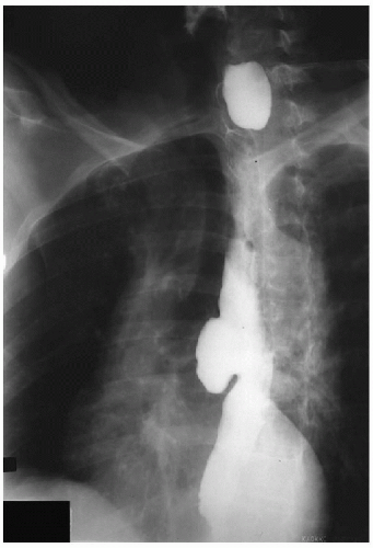
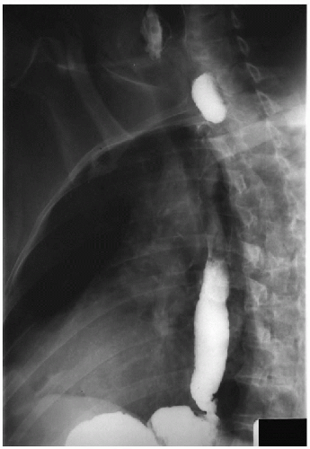
▪ Esophagogram
Esophagogram Report
The esophagograms seen demonstrate a diverticulum in the proximal esophagus, which remains filled with contrast after most of the contrast has cleared from the esophagus. This finding is characteristic for a Zenker’s diverticulum. ▪


