Wound Healing

Chapter 7 Wound Healing


The treatment and healing of wounds are some of the oldest subjects discussed in the medical literature. Yet despite knowledge of the steps involved, the exact mechanisms underlying wound healing are not completely understood.


Wound-Healing Phases


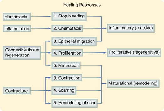
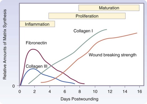
The three phases of wound healing are inflammation, proliferation, and maturation. In a large wound such as a pressure sore, the eschar or fibrinous exudate reflects the inflammatory phase, the granulation tissue is part of the proliferative phase, and the contracting or advancing edge is part of the maturational phase. All three phases may occur simultaneously, and the phases may overlap with their individual processes (Fig. 7-1).


Inflammatory Phase


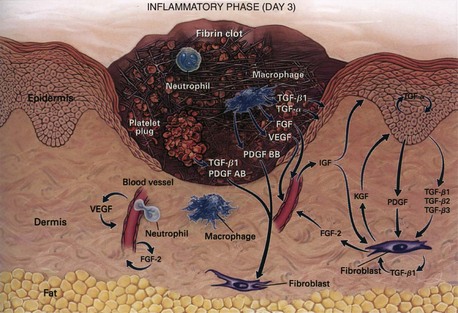
During the immediate reaction of the tissue to injury, hemostasis and inflammation occur. This phase represents an attempt to limit damage by stopping the bleeding, sealing the surface of the wound, and removing any necrotic tissue, foreign debris, or bacteria present. The inflammatory phase is characterized by increased vascular permeability, migration of cells into the wound by chemotaxis, secretion of cytokines and growth factors into the wound, and activation of the migrating cells (Fig. 7-2).


Hemostasis and Inflammation


During an acute tissue injury, blood vessel damage results in initial intense local vasoconstriction of arterioles and capillaries followed by vasodilation and increased vascular permeability (Fig. 7-3). Erythrocytes and platelets adhere to the damaged capillary endothelium, resulting in plugging of capillaries and leading to cessation of hemorrhage. Activation of these platelets by binding to the exposed type IV and V collagen from the damaged endothelium results in platelet aggregation. The initial contact between platelets and collagen requires von Willebrand factor (vWF) VIII, a heterodimeric protein synthesized by megakaryocytes and endothelial cells. Platelet adhesion to the endothelium is primarily mediated through the interaction between high-affinity glycoprotein receptors and the integrin receptor GPIIb-IIIa (αIIbβ3). Platelets also express other integrin receptors that mediate direct binding to collagen (α2β1) and laminin (α6β1) or indirect binding by attaching to subendothelial matrix-bound fibronectin (α5β1), vitronectin (αvβ3), and other ligands.


Increased Vascular Permeability


Platelet binding results in conformational changes in platelets that trigger intracellular signal transduction pathways that lead to platelet activation and the release of biologically active proteins. Platelet alpha granules are storage organelles that contain platelet-derived growth factor (PDGF), transforming growth factor-β (TGF-β), insulin-like growth factor type I (IGF-I), fibronectin, fibrinogen, thrombospondin, and vWF. The dense bodies contain vasoactive amines, such as serotonin, that cause vasodilation and increased vascular permeability. Mast cells adherent to the endothelial surface release histamine and serotonin, resulting in increased permeability of endothelial cells and causing leakage of plasma from the intravascular space to the extracellular compartment. The clotting cascade is initiated through the intrinsic and extrinsic pathways. As the platelets become activated, the membrane phospholipids bind factor V, which allows interaction with factor X. Membrane-bound prothrombinase activity is generated and potentiates thrombin production exponentially. The thrombin itself activates platelets and catalyzes the conversion of fibrinogen to fibrin. The fibrin strands trap red blood cells to form the clot and seal the wound. The lattice framework that results will be the scaffold for endothelial cells, inflammatory cells, and fibroblasts. Thromboxane A2 and prostaglandin F2α, formed from the degradation of cell membranes in the arachidonic acid cascade, also assist in platelet aggregation and vasoconstriction. Although these activities serve to limit the amount of injury, they can also cause localized ischemia, resulting in further damage to cell membranes and the release of more prostaglandin F2α and thromboxane A2.


Chemokines


Chemokines stimulate the migration of different cell types, particularly inflammatory cells, into the wound and are active participants in the regulation of the different phases of wound healing. The CXC, CC, and C ligand families bind to G protein–coupled surface receptors called CXC receptors and CC receptors.


Macrophage chemoattractant protein (MCP-1, or CCL2) is induced in keratinocytes after injury. It is a potent chemoattractant for monocytes/macrophages, T lymphocytes, and mast cells.1 Expression of this chemokine is sustained in chronic wounds and results in the prolonged presence of polymorphonuclear cells (PMNs) and macrophages, leading to the prolonged inflammatory response.2 CXCL1 (GRO-α) is a potent PMN chemotactic regulator and is increased in acute wounds. It is also involved in reepithelialization.3 Interleukin-8 (IL-8, or CXCL8) expression is increased in acute and chronic wounds.4 It is involved in reepithelialization and induces the leukocyte expression of matrix metalloproteinases (MMPs), which stimulates remodeling. It is also a strong chemoattractant for PMNs and participates in inflammation.4 Relatively low levels of IL-8 are found in fetal wounds and may be why fetal wounds have decreased inflammation and heal without scars.5 Expression of the keratinocyte-produced chemokine interferon inducible protein 10 (IP-10 or CXCL10) is elevated in acute wounds as well as chronic inflammatory conditions.6 It impairs wound healing by increasing inflammation and recruiting lymphocytes to the wound. It also inhibits proliferation by decreasing reepithelialization and angiogenesis and preventing fibroblast migration.3 Stromal cell-derived factor-1 (SDF-1, or CXCL12) is expressed by endothelial cells, myofibroblasts, and keratinocytes and is involved in inflammation by recruiting lymphocytes to the wound and promoting angiogenesis. It is a potent chemoattractant for endothelial cells and bone marrow progenitors from the circulation to peripheral tissues.7 It also enhances keratinocyte proliferation, resulting in reepithelialization.8


Polymorphonuclear Cells


The release of histamine and serotonin leads to vascular permeability of the capillary bed. Complement factors such as C5a and leukotriene B4 promote neutrophil adherence and chemoattraction. In the presence of thrombin, endothelial cells exposed to leukotriene C4 and D4 release platelet-aggregating factor, which further enhances neutrophil adhesion. Monocytes and endothelial cells produce the inflammatory mediators IL-1 and tumor necrosis factor-α (TNF-α), and these mediators further promote endothelial-neutrophil adherence. The increased capillary permeability and the various chemotactic factors facilitate diapedesis of neutrophils into the inflammatory site. As the neutrophils begin their migration, they release the contents of their lysosomes and enzymes such as elastase and other proteases into the extracellular matrix (ECM), which further facilitates neutrophil migration. The combination of intense vasodilation and increased vascular permeability leads to clinical findings of inflammation, rubor (redness), tumor (swelling), calor (heat), and dolor (pain). Local tissue swelling is further promoted by the deposition of fibrin, a protein end product of coagulation, and the fibrin becomes entrapped in lymphatic vessels.


Evidence suggests that the migration of PMNs requires sequential adhesive and de-adhesive interactions between β1 and β2 integrins and ECM components. Integrin molecules are a family of cell surface receptors that are closely coupled with the cell’s cytoskeleton. These molecules serve two major functions:


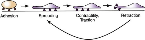
Integrins are crucial for cell motility and are required in inflammation and normal wound healing, as well as in embryonic development and tumor metastases. After extravasation, PMNs, attracted by chemotaxins, migrate through the ECM by means of transient interactions between integrin receptors and their ligands. Four phases of integrin-mediated cell motility have been described: adhesion, spreading, contractility or traction, and retraction. Activation of specific integrins though ligand binding has been shown to increase cell adhesion and activate reorganization of the cell’s actin cytoskeleton. Spreading is characterized by the development of lamellipodia and filopodia. Traction at the leading edge of the cell develops through binding of integrin, followed by translocation of the cell over the adherent segment of the plasma membrane. The integrin is shifted to the rear of the cell and releases its substrate, thereby permitting cell advancement (Fig. 7-4). Regulation of integrin function by adhesive substrates offers a mechanism for local control of migrant cells. Within the assembled framework of the ECM, binding sites for integrins have been identified on collagen, laminin, and fibronectin.


The chemotactic agent mediates the PMN response through signal transduction as the chemotaxin binds to receptors on the cell surface. Bacterial products such as N-formyl-methionyl-leucyl-phenylalanine bind to induce cyclic adenosine monophosphate (cAMP) but if there is maximal receptor occupancy, superoxide is produced at peak rates. Neutrophils also possess receptors for IgG and the complement proteins C3b and C3bi. As the complement cascade is released and bacteria are opsonized, binding of these proteins to cell receptors on neutrophils allows recognition by the neutrophils and phagocytosis of the bacteria. When neutrophils are stimulated, they express more CR1 and CR3 receptors, thereby permitting more efficient binding and phagocytosis of these bacteria.


Functional activation occurs after migration of PMNs into the wound site, which may induce new cell surface antigen expression, increased cytotoxicity, or enhanced production and release of cytokines. These activated neutrophils scavenge for necrotic debris, foreign material, and bacteria and generate free oxygen radicals, with electrons donated by the reduced form of nicotinamide adenine dinucleotide phosphate (NADPH). The electrons are transported across the membrane into lysosomes, where superoxide anion (O2) is formed. Superoxide dismutase catalyzes the formation of hydrogen peroxide (H2O2), which is then degraded by myeloperoxidase in the azurophilic granules of neutrophils. This interaction oxidizes halides, with the formation of byproducts such as hypochlorous acid. The iron-catalyzed reaction between H2O2 and O2 forms hydroxyl radicals (OH·). This potent free radical is bactericidal, but is also toxic to neutrophils and surrounding viable tissues.


Migration of PMNs stops when wound contamination has been controlled, usually within the first few days after injury. PMNs do not survive longer than 24 hours. After 24 to 48 hours, the predominance of cells in the wound cleft shifts to mononuclear cells. If wound contamination persists or secondary infection occurs, continuous activation of the complement system and other pathways provides a steady supply of chemotactic factors, resulting in a sustained influx of PMNs into the wound. In addition to the delay in healing, this prolonged inflammation can be deleterious in terms of destruction of normal tissue, with progression to tissue necrosis, abscess formation, and possibly systemic infection. PMNs are not essential for wound healing because their role in phagocytosis and antimicrobial defense may be taken over by macrophages. Sterile incisions will heal normally without the presence of PMNs.


Macrophages


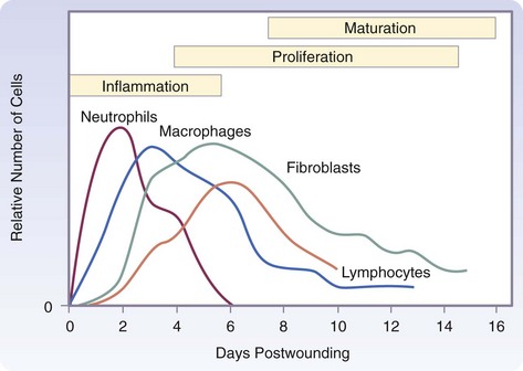
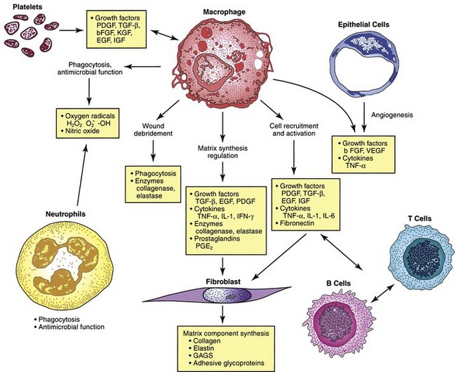
The macrophage is the one cell that is truly crucial to wound healing in that it serves to orchestrate the release of cytokines and stimulate many of the subsequent processes of wound healing (Fig. 7-5). Macrophages in the wound appear at the same time that neutrophils disappear. Macrophages induce apoptosis of PMNs. Chemotaxis of migrating blood monocytes occurs within 24 to 48 hours. Chemotactic factors specific for monocytes include bacterial products, complement degradation products (C5a), thrombin, fibronectin, collagen, TGF-β, and PDGF-BB. Monocyte chemotaxis is also facilitated by the interaction of integrin receptors on the monocyte surface with ECM proteins such as fibrin and fibronectin. The β integrin receptor also transduces the signal for macrophage phagocytic activity. Activated integrin expression promotes adhesion-mediated gene induction in monocytes that transforms them into wound macrophages; such transformation results in increased phagocytic activity and selective expression of cytokines and signal transduction elements by messenger RNA (mRNA), including the early growth response genes EGR2 and c-fos. Macrophages have specific receptors for immunoglobulin G (IgG; Fc receptor), C3b (CR1 and CR3), and fibronectin (integrin receptors) that permit surface recognition of opsonized pathogens and facilitate phagocytosis.


Bacterial debris such as lipopolysaccharide can activate monocytes to release free radicals and cytokines that mediate angiogenesis and fibroplasia. The presence of IL-2 increases the release of free radicals and thus enhances bactericidal activity, and the activity of the free radicals is potentiated by IL-2. In addition, the free radicals generate bacterial debris, which further potentiates the activation of monocytes. Activated wound macrophages also produce nitric oxide (NO), a substance that has been demonstrated to have many functions other than antimicrobial properties.


As the monocyte or macrophage is activated, phospholipase is induced, cell membrane phospholipids are enzymatically degraded, and thromboxane A2 and prostaglandin F2α are released. The macrophage also releases leukotrienes B4 and C4 and 15- and 5-hydroxyeicosatetraenoic acid. Leukotriene B4 is a potent chemotaxin for neutrophils and increases their adherence to endothelial cells.


Wound macrophages release proteinases, including matrix metalloproteinases (MMP-1, MMP-2, MMP-3, and MMP-9; Fig. 7-6), that degrade the ECM and are crucial for removing foreign material, promoting cell movement through tissue spaces, and regulating ECM turnover. This activity is dependent on the cAMP pathway and thus can be blocked by nonsteroidal anti-inflammatory drtugs (NSAIDs) or glucocorticoid drugs. Colchicine and retinoic acid appear to decrease collagenase production as well.


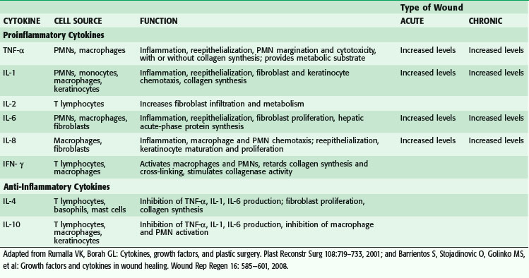
Macrophages secrete numerous cytokines and growth factors (Tables 7-1 and 7-2). IL-1, a proinflammatory cytokine, is an acute-phase response cytokine. This endogenous pyrogen causes lymphocyte activation and stimulation of the hypothalamus, thereby inducing the febrile response. It also directly affects hemostasis by inducing the release of vasodilators and stimulating coagulation. Its effect is further amplified as endothelial cells produce it in the presence of TNF-α and endotoxin. IL-1 has numerous effects, such as enhancement of collagenase production, stimulation of cartilage degradation and bone reabsorption, activation of neutrophils, regulation of adhesion molecules, and promotion of chemotaxis. It stimulates other cells to secrete proinflammatory cytokines. Its effects also extend into the proliferative phase, during which it increases fibroblast and keratinocyte growth and collagen synthesis. Studies have demonstrated increased levels of IL-1 in chronic nonhealing wounds, thus suggesting its role in the pathogenesis of poor wound healing. The early beneficial responses of IL-1 in wound healing appear to be maladaptive if elevated levels last beyond the first week after injury.


Microbial byproducts induce macrophages to release TNF. TNF-α is crucial in initiating the response to injury or bacteria. It upregulates cell surface adhesion molecules that promote the interaction of immune cells and endothelium. TNF-α is detected in the wound within 12 hours and peaks after 72 hours. Its effects include hemostasis, increased vascular permeability, and enhanced endothelial proliferation. Like IL-1, TNF-α induces fever, increased collagenase production, reabsorption of cartilage and bone, and release of PDGF, as well as the production of more IL-1. Excessive production of TNF-α, however, has been associated with multisystem organ failure and increased morbidity and mortality in inflammatory disease states, partly through its effects on activating macrophages and neutrophils. Studies have noted elevated levels of TNF-α in nonhealing versus healing chronic venous ulcers. Thus, as in the case of IL-1, TNF-α appears to be essential in the early inflammatory response required for wound healing, but local and systemic persistence of this cytokine may lead to impaired wound maturation.


IL-6, which is produced by monocytes and macrophages, is involved in stem cell growth, activation of B and T cells, and regulation of the synthesis of hepatic acute-phase proteins. Within acute wounds, IL-6 is also secreted by PMNs and fibroblasts, and its increase parallels the increase in the PMN count locally. IL-6 is detectable within 12 hours of experimental wounding and may persist at high concentrations for longer than 1 week. It also works synergistically with IL-1, TNF-α, and endotoxins. It is a potent stimulator of fibroblast proliferation and is decreased in aging fibroblasts and fetal wounds.


IL-8 (also called CXCL8) is secreted primarily by macrophages and fibroblasts in the acute wound, with peak expression within the first 24 hours. Its major effects have been discussed but include increased PMN and monocyte chemotaxis, PMN degranulation, and expression of endothelial cell adhesion molecules.


Interferon-γ (IFN-γ), another proinflammatory cytokine, is secreted by T lymphocytes and macrophages. Its major effects are macrophage and PMN activation and increased cytotoxicity. It has also been shown to reduce local wound contraction and aid in tissue remodeling. IFN-γ has been used in the treatment of hypertrophic and keloid scars, possibly by its effect in slowing collagen production and cross linking, whereas collagenase (MMP-1) production increases. Experimentally, however, it has been shown to impair reepithelialization and wound strength in a dose-dependent manner when applied locally or systemically. These findings suggest that administration of IFN-γ may improve scar hypertrophy by decreasing the strength of the wound.


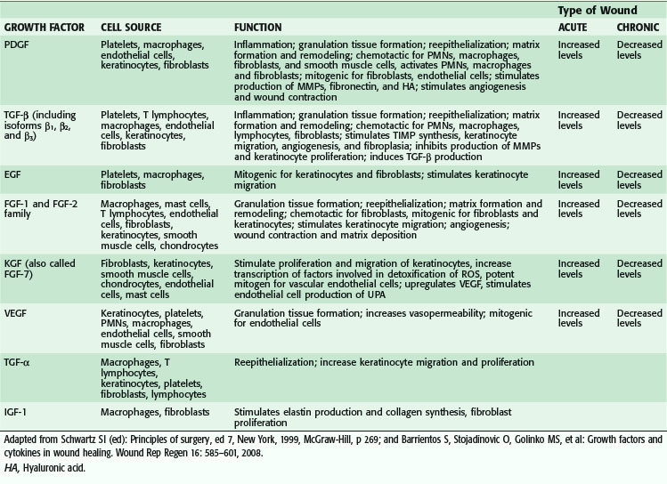
Macrophages also release growth factors that stimulate fibroblast, endothelial cell, and keratinocyte proliferation and are important in the proliferative phase (see Table 7-2). Macrophage-secreted PDGF stimulates collagen and proteoglycan synthesis. PDGF exists as three isomers—PDGF-AA, PDGF-AB, and PDGF-BB. However, the PDGF-BB isomer is the only growth factor preparation approved by the U.S. Food and Drug Administration and is the most widely studied clinically. Topical application of recombinant PDGF has improved wound-breaking strength and healing time in human and murine models of acute wounding. Administration of PDGF-BB has improved wound closure in chronic and diabetic nonhealing ulcers in humans and rodents but did not have the same effect in steroid-treated animals.


TGF-α and TGF-β are both released by activated monocytes. TGF-α stimulates epidermal growth and angiogenesis. TGF-β itself stimulates monocytes to express other peptides such as TGF-α, IL-1, and PDGF. TGF-β, which is also released by platelets and fibroblasts within wounds, exists as at least three isomers—β1, β2, and β3—and its effects include fibroblast migration and maturation and ECM synthesis. TGF-β1 has been shown to play an important role in collagen metabolism and healing of gastrointestinal injuries and anastomoses. In experimental models, TGF-β1 accelerates wound healing in normal, steroid-impaired, and irradiated animals.


TGF-β is the most potent stimulant of fibroplasia, and its strong mitogenic effects have been implicated in the fibrogenesis seen in disease states such as scleroderma and interstitial pulmonary fibrosis. Enhanced expression of TGF-β1 mRNA is found in keloid and hypertrophic scars. In contrast, fetal wounds have been demonstrated to have a paucity of TGF-β, thus suggesting that the scarless repair seen in utero occurs because of low or absent amounts of TGF-β. Studies of the three isomers have suggested that although TGF-β1 and TGF-β2 play an important role in tissue fibrosis and postinjury scarring, TGF-β3 may limit scarring. As the concentration of TGF-β rises in the inflammatory site, fibroblasts are directly stimulated to produce collagen and fibronectin, thus leading to the proliferative phase.


Lymphocytes


T lymphocytes appear in significant number in the wound at approximately the fifth day, with a peak occurring at approximately the seventh day. B lymphocytes do not appear to play a significant role in wound healing but seem to be involved in downregulating healing as the wound closes. Lymphocytes exert most of their effects on fibroblasts by producing stimulatory cytokines, such as IL-2 and fibroblast-activating factor, and inhibitory cytokines, such as TGF-β, TNF-α, and IFN-γ. Initially, lymphocytes were thought to play a minimal role in acute wound healing, particularly in the absence of excessive inflammation. The macrophage processes foreign debris such as bacteria or enzymatically degraded host proteins and serves as an antigen-presenting cell to lymphocytes. This interaction stimulates lymphocyte proliferation and release of cytokines. T cells produce IFN-γ, which stimulates the macrophage to release a cascade of cytokines, including TNF-α and IL-1. IFN-γ also causes decreased synthesis of prostaglandins, which enhances the effect of inflammatory mediators. In addition, IFN-γ suppresses collagen synthesis and inhibits macrophages from leaving the site of injury. Thus, IFN-γ appears to be an important mediator of chronic nonhealing wounds, and its presence suggests that T lymphocytes are primarily involved in chronic wound healing.


Some studies, however, have questioned the belief that lymphocytes are not essential for acute wound healing. Drugs that suppress T-lymphocyte function and proliferation, such as steroids and immunosuppressive agents (e.g., cyclosporine, tacrolimus), have been found to result in impaired wound healing in experimental wound models, possibly through decreased NO synthesis. In vivo lymphocyte depletion suggests the existence of an incompletely characterized T cell lymphocyte population that is neither CD4+ nor CD8+, and it is this subset that seems to be responsible for the promotion of wound healing.


Proliferative Phase


As the acute responses of hemostasis and inflammation begin to resolve, the scaffolding is laid for repair of the wound through angiogenesis, fibroplasia, and epithelialization. This stage is characterized by the formation of granulation tissue, which consists of a capillary bed, fibroblasts, macrophages, and a loose arrangement of collagen, fibronectin, and hyaluronic acid.


A number of studies have used growth factors to modify granulation tissue, particularly fibroplasia. Adenoviral transfer, topical application, and subcutaneous injection of PDGF, TGF-β, keratinocyte growth factor (KGF), vascular endothelial growth factor (VEGF), and epidermal growth factor (EGF) have been tested to increase the proliferation of granulation tissue.


Angiogenesis


Angiogenesis is the process of new blood vessel formation and is necessary to support a healing wound environment. After injury, activated endothelial cells degrade the basement membrane of postcapillary venules, thereby allowing the migration of cells through this gap. Division of these migrating endothelial cells results in tubule or lumen formation. Eventually, deposition of the basement membrane occurs and results in capillary maturation.


After injury, the endothelium is exposed to numerous soluble factors and comes in contact with adhering blood cells. These interactions result in upregulation of the expression of cell surface adhesion molecules, such as vascular cell surface adhesion molecule-1 (VCAM-1). Matrix-degrading enzymes, such as plasmin and the metalloproteinases, are released and activated, and degrade the endothelial basement membrane. Fragmentation of the basement membrane allows migration of endothelial cells into the wound, promoted by fibroblast growth factor (FGF), PDGF, and TGF-β. Injured endothelial cells express adhesion molecules, such as the integrin αvβ3, which facilitates attachment to fibrin, fibronectin, and fibrinogen and thus facilitates endothelial cell migration along the provisional matrix scaffold. Platelet endothelial cell adhesion molecule-1 (PECAM-1), also found on endothelial cells, modulates their interaction with each other as they migrate into the wound.


Capillary tube formation is a complex process that involves cell-cell and cell-matrix interactions, modulated by adhesion molecules on endothelial cell surfaces. PECAM-1 has been observed to mediate cell-cell contact, whereas β1 integrin receptors may aid in stabilizing these contacts and forming tight junctions between endothelial cells. Some of the new capillaries differentiate into arterioles and venules, whereas others undergo involution and apoptosis, with subsequent ingestion by macrophages. Regulation of endothelial apoptosis is not well understood.


Angiogenesis appears to be stimulated and manipulated by a variety of cytokines predominantly produced by macrophages and platelets. As the macrophage produces TNF-α, it orchestrates angiogenesis during the inflammatory phase. Heparin, which can stimulate the migration of capillary endothelial cells, binds with high affinity to a group of angiogenic factors.


VEGF, a member of the PDGF family of growth factors, has potent angiogenic activity. It is produced in large amounts by keratinocytes, macrophages, endothelial cells, platelets, and fibroblasts during wound healing.912 Cell disruption and hypoxia, hallmarks of tissue injury, appear to be strong initial inducers of potent angiogenic factors at the wound site, such as VEGF and its receptor. VEGF family members include VEGF-A, VEGF-B, VEGF-C, VEGF-D, VEGF-E, and placental growth factor (PLGF).13 VEGF-A promotes early events in angiogenesis and subsequently is crucial to wound healing.14 It binds to tyrosine kinase surface receptors Flt-1 (VEGF receptor-1, or VEGFR-1) and KDR (VEGF receptor-2, or VEGFR-2).15 Flt-1 is required for blood vessel organization, whereas KDR is important for endothelial cell chemotaxis, proliferation, and differentiation.16,17 Animal studies have shown that VEGF-A administration restores impaired angiogenesis found in diabetic ischemic limbs18; however, other studies have shown that exogenous VEGF results in vascular leakage and disorganized blood vessel formation.19,20 VEGF-C, which is also elevated during wound healing, is primarily released by macrophages and is important during the inflammatory phase of wound healing.21 Although it works primarily through VEGF receptor-3 (VEGFR-3), which is expressed in macrophages and lymphatic endothelium, it can also activate VEGFR-2, thereby increasing vascular permeability.21 In vivo administration of VEGF-C in an animal model using an adenoviral vector to genetically diabetic mice resulted in accelerated healing.13 PLGF is another proangiogenic factor that is elevated after wounding. It is involved in inflammation and expressed by keratinocytes and endothelial cells. It is believed to work synergistically with VEGF, thereby potentiating its proangiogenic function.22


Both acidic and basic FGFs (FGF-1 and FGF-2) are released from disrupted parenchymal cells and are early stimulants of angiogenesis. FGF-2 provides the initial angiogenic stimulus within the first 3 days of wound repair, followed by a subsequent prolonged stimulus mediated by VEGF from days 4 through 7. There is a dose-dependent effect of VEGF and FGF-2 on angiogenesis. Both TGF-α and EGF stimulate endothelial cell proliferation. TNF-α is chemotactic for endothelial cells; it promotes formation of the capillary tube and may mediate angiogenesis through its induction of hypoxia-inducible factor 1 (HIF-1). It regulates the expression of other hypoxia-responsive genes, including inducible NO synthase and VEGF. HIF-1α mRNA is prominently present in wound inflammatory cells during the initial 24 hours, and HIF-1α protein is present in cells isolated from the wound 1 and 5 days after injury in vitro. Data also suggest that there is a positive interaction between endogenous NO and VEGF, with endogenous NO enhancing VEGF synthesis. Similarly, VEGF has been shown to promote NO synthesis in angiogenesis, thus suggesting that NO mediates aspects of VEGF signaling required for endothelial cell proliferation and organization.


TGF-β is a chemoattractant for fibroblasts and probably assists in angiogenesis by signaling the fibroblast to produce FGFs. Other factors that have been shown to induce angiogenesis include angiogenin, IL-8, and lactic acid. Several of the matrix materials, such as fibronectin and hyaluronic acid from the wound site, are angiogenic. Fibronectin and fibrin are produced by macrophages and damaged endothelial cells. Collagen appears to interact by causing the tubular formation of endothelial cells in vitro. Angiogenesis thus results from the complex interaction of ECM material and cytokines.


Fibroplasia


Fibroblasts are specialized cells that differentiate from resting mesenchymal cells in connective tissue; they do not arrive in the wound cleft by diapedesis from circulating cells. After injury, the normally quiescent and sparse fibroblasts are chemoattracted to the inflammatory site, where they divide and produce the components of the ECM. After stimulation by macrophage- and platelet-derived cytokines and growth factors, the fibroblast, which is normally arrested in the G0 phase, undergoes replication and proliferation. Platelet-derived TGF-β stimulates fibroblast proliferation indirectly by releasing PDGF. The fibroblast can also stimulate replication in an autocrine manner by releasing FGF-2. To continue proliferating, fibroblasts require further stimulation by factors such as EGF or IGF-I. Although fibroblasts require growth factors for proliferation, they do not need growth factors to survive. Fibroblasts can live quiescently in growth factor–free media in monolayers or three-dimensional cultures.


The primary function of fibroblasts is to synthesize collagen, which they begin to produce during the cellular phase of inflammation. The time required for undifferentiated mesenchymal cells to differentiate into highly specialized fibroblasts accounts for the delay between injury and the appearance of collagen in a healing wound. This period, generally 3 to 5 days, depending on the type of tissue injured, is called the lag phase of wound healing. Fibroblasts begin to migrate in response to chemotactic substances such as growth factors (PDGF, TGF-β), C5 fragments, thrombin, TNF-α, eicosanoids, elastin fragments, leukotriene B4, and fragments of collagen and fibronectin.


The rate of collagen synthesis declines after 4 weeks and eventually balances the rate of collagen destruction by collagenase (MMP-1). At this point, the wound enters a phase of collagen maturation. The maturation phase continues for months or even years. Glycoprotein and mucopolysaccharide levels decrease during the maturation phase, and new capillaries regress and disappear. These changes alter the appearance of the wound and increase its strength.


Epithelialization


The epidermis serves as a physical barrier to prevent fluid loss and bacterial invasion. Tight cell junctions within the epithelium contribute to its impermeability and the basement membrane zone gives structural support and provides attachment between the epidermis and the dermis. The basement membrane zone consists of several layers:


The basal layer of the epidermis attaches to the basement membrane zone by hemidesmosomes. Reepithelialization of wounds begins within hours after injury. Initially, the wound is rapidly sealed by clot formation and then by epithelial (epidermal) cell migration across the defect. Keratinocytes located at the basal layer of the residual epidermis or in the depths of epithelium-lined dermal appendages migrate to resurface the wound. Epithelialization involves a sequence of changes in wound keratinocytes—detachment, migration, proliferation, differentiation, and stratification. If the basement membrane zone is intact, epithelialization proceeds more rapidly. The cells are stimulated to migrate. Attachments to neighboring and adjoining cells and to the dermis are loosened, as demonstrated by intracellular tonofilament retraction, dissolution of intercellular desmosomes and hemidesmosomes linking the epidermis to the basement membrane, and formation of cytoplasmic actin filaments.


Epidermal cells express integrin receptors that allow them to interact with ECM proteins such as fibronectin. The migrating cells dissect the wound by separating the desiccated eschar from viable tissue. This path of dissection is determined by the integrins that the epidermal cells express on their cell membranes. Degradation of the ECM, required if epidermal cells are to migrate between the collagenous dermis and fibrin eschar, is driven by epidermal cell production of collagenase (MMP-1) and plasminogen activator, which activates collagenase and plasmin. The migrating cells are also phagocytic and remove debris in their path. Cells behind the leading edge of migrating cells begin to proliferate. The epithelial cells move in a leapfrog and tumbling fashion until the edges establish contact. If the basement membrane zone is not intact, it will be repaired first. The absence of neighboring cells at the wound margin may be a signal for the migration and proliferation of epidermal cells. Local release of EGF, TGF-α, and KGF and increased expression of their receptors may also stimulate these processes. Topical application of KGF-2 in young and aged animals accelerates reepithelialization. Basement membrane proteins, such as laminin, reappear in a highly ordered sequence from the margin of the wound inward. After the wound is completely reepithelialized, the cells become columnar and stratified again while firmly attaching to the reestablished basement membrane and underlying dermis.


Extracellular Matrix


The ECM exists as a scaffold to stabilize the physical structure of tissues, but it also plays an active and complex role by regulating the behavior of cells that contact it. Cells within it produce the macromolecular constituents, including the following:


In connective tissue, proteoglycan molecules form a gel-like ground substance. This highly hydrated gel allows the matrix to withstand compressive force while permitting rapid diffusion of nutrients, metabolites, and hormones between blood and tissue cells. Collagen fibers within the matrix serve to organize and strengthen it, whereas elastin fibers give it resilience and matrix proteins have adhesive functions.23


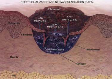
The wound matrix accumulates and changes in composition as healing progresses, balanced between new deposition and degradation (Fig. 7-7). The provisional matrix is a scaffold for cellular migration and is composed of fibrin, fibrinogen, fibronectin, and vitronectin. GAGs and proteoglycans are synthesized next and support further matrix deposition and remodeling. Collagens, which are the predominant scar proteins, are the end result. Attachment proteins, such as fibrin and fibronectin, provide linkage to the ECM through binding to cell surface integrin receptors.


Stimulation of fibroblasts by growth factors induces upregulated expression of integrin receptors, thereby facilitating cell-matrix interactions. Ligand binding induces clustering of integrin into focal adhesion sites. Regulation of integrin-mediated cell signaling by the extracellular divalent cations Mg2+, Mn2+, and Ca2+ is perhaps caused by induction of conformational changes in the integrins.


A dynamic and reciprocal relationship exists between fibroblasts and the ECM. Cytokine regulation of fibroblast responses is altered by variations in the composition of the ECM. For example, expression of matrix-degrading enzymes, such as the MMPs, is upregulated after cytokine stimulation of fibroblasts. Collagenolytic MMP-1 is induced by IL-1 and downregulated by TGF-β. Activation of plasminogen to plasmin by plasminogen activator and procollagenase to collagenase by plasmin results in matrix degradation and facilitates cell migration. Modulation of these processes provides additional mechanisms whereby the cell-matrix interaction can be regulated during wound healing. Matrix modulation is also seen in tumor metastasis. Neoplastic cells lose their dependence on anchorage, mediated mainly by integrins; this is probably caused by decreased production of fibronectin and subsequent decreased adhesion and, as a result, these cells can break away from the primary tumor and metastasize.


An example of the necessary dynamic interactions occurring in the provisional matrix during wound healing is the effect of TGF-β on incisional wounds sealed with fibrin sealant. Fibrin sealant is a derivative of plasma components that mimics the last step in the coagulation cascade. Commercially available fibrin sealant has an approximately 10-fold greater concentration of fibrin than plasma and consequently provides a more airtight, waterproof seal. Fibrin sealant may serve as a mechanical barrier to the early cell-mediated events occurring in wound healing. Supplementation of fibrin sealant with TGF-β has been demonstrated to reverse the inhibitory effects of fibrin sealant on wound healing and increase tensile strength as compared with sutured wounds. The increased tensile strength may be a result of improved cell migration into the wound site, more rapid clearance of fibrin sealant, suppression of gelatinase (MMP-9), and enhancement of ECM synthesis in TGF-β–supplemented wounds.


Collagen Structure


Collagens are found in all multicellular animals and are secreted by a variety of cell types. They are a major component of skin and bone and constitute 25% of the total protein mass in mammals. The proline- and glycine-rich collagen molecule is a long, stiff, triple-stranded helical structure that consists of three collagen polypeptide α chains wound around one another in a ropelike superhelix. With its ringlike structure, proline provides stability to the helical conformation in each α chain, whereas glycine, because of its small size, allows tight packing of the three α chains to form the final superhelix. There are at least 20 types of collagen, the main constituents of connective tissue being types I, II, III, V, and XI. Type I is the principal collagen of skin and bone and is the most common.23 In adults, the skin is approximately 80% type I and 20% type III. In newborns, the content of type III collagen is greater than that found in adults. In early wound healing, there is also increased expression of type III collagen. Type I collagens are the fibrillar, or fibril-forming, collagens. They are secreted into the extracellular space, where they assemble into collagen fibrils (10 to 300 nm in diameter), which then aggregate into larger, cable-like bundles called collagen fibers (several micrometers in diameter).


Other types of collagens include types IX and XII (fibril-associated collagens) and types IV and VII (network-forming collagens). Types IX and XII are found on the surface of collagen fibrils and serve to link the fibrils to one another and to other components in the ECM. Type IV molecules assemble into a meshlike pattern and are a major part of the mature basal lamina. Dimers of type VII form anchoring fibrils that help attach the basal lamina to the underlying connective tissue and are especially abundant in the skin.


Type XVII and XVIII collagens are two of a number of collagen-like proteins. Type XVII has a transmembrane domain and is found in hemidesmosomes. Type XVIII is located in the basal laminae of blood vessels. The peptide endostatin, which inhibits angiogenesis and shows promise as an anticancer drug, is formed by cleavage of the C-terminal domain of type XVIII collagen.

< div class='tao-gold-member'>

Stay updated, free articles. Join our Telegram channel

Aug 1, 2016 | Posted by in CARDIAC SURGERY | Comments Off on Wound Healing

Full access? Get Clinical Tree

Get Clinical Tree app for offline access