Fig. 7.1
Cardiovascular disease in adults aged 20 years or older by age and sex. The prevalence of coronary artery disease (CAD) in women and men increases as they age. Younger women (aged 20–39 years) have a much lower risk of CAD compared with men. However, by age 60, women catch up with their male counterparts and by age 80 the percentage of women with CAD exceeds that of men (Adapted from National Institutes of Health; National Heart, Lung, and Blood Institute [1])
Cardiovascular Disease: Women and Ethnic Background
As the population of ethnically diverse persons increases in the United States, it becomes equally important to understand cardiovascular risk factors in these groups in order to treat them appropriately. Although most physicians acknowledge the racial and ethnic disparities in the delivery of health care, they are not likely to recognize this imbalance within their own clinical practice [7]. Minorities have been shown to have a higher burden of cardiovascular disease and increased morbidity, reduced quality of life, and death. For example, the rate of death due to cardiovascular causes is higher in black women than in white women [8]. There is a growing database on disease differences in blacks and Hispanics, but the data on Asians continue to be scarce and many health differences remain unknown [9].
Ethnic minority patients with cardiovascular disease tend to be younger, to have many more comorbidities such as diabetes mellitus and hypertension, and to be female [10]. Compared with white patients, minority patients who present with acute coronary syndrome are less likely to undergo angiography [11]. In addition, despite the prevalence of known risk factors for cardiovascular disease, such as hypertension and diabetes, black women tend to receive suboptimal treatment for these diseases [12]. Hispanics undergoing coronary artery bypass grafting tend to be younger and are more likely to be female, with a lower body mass index than that of other patients [13]. Some differences in treatment and delays in presentation can be attributed to a lack of awareness; however, these differences may also be mediated by the patient’s race or sex, and/or the physician’s perceptions [14].
Cardiovascular Risk Assessment: Risk Engines
Several models have been proposed to predict cardiovascular risk. Perhaps the most utilized is the Framingham risk score (Fig. 7.2) [15]. The Framingham risk score has been criticized for representing a small subset of the overall population of women. The study mainly included white middle-class women, thereby perhaps making its applicability to minority women less cogent. The Framingham risk score does not take into account smoking history, body mass index (height in m2/weight in kg), exercise capacity or level of fitness. Furthermore, it should be noted that even up to age 80 years, most women are considered to be at low risk despite an increased lifetime risk.
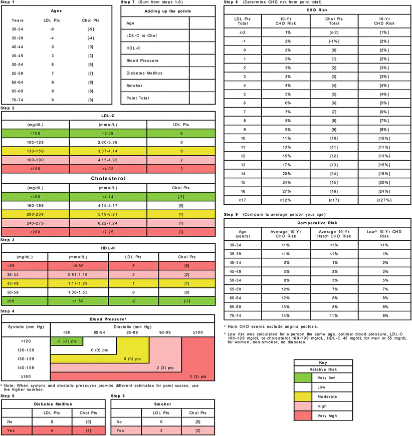
Fig. 7.2
Framingham risk score. This score estimates the risk of coronary heart disease (CHD) over a 10-year period based on the Framingham experience. Chol indicates cholesterol, HDL-C high-density lipoprotein cholesterol, LDL low-density lipoprotein, LDL-C low-density lipoprotein cholesterol, Pts points, yr year (Adapted from Wilson et al. [15]. Used with permission from Wolters Kluwer Health)
Figure 7.3 [16] demonstrates risk reassignment using the Reynolds risk score. Briefly, this score incorporates high-sensitivity C-reactive protein, hemoglobin A1c in the presence of diabetes, and family history. This score assesses the 10-year global cardiovascular risk perhaps more accurately than does the Framingham score. Both are recommended in the American College of Cardiology/American Heart Association (ACC/AHA) guidelines as tools to risk-stratify patients. However, risk factor modifications have a differential effect on cardiovascular risk for men and women [17].
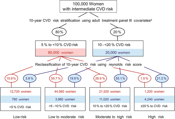
Fig. 7.3
Reynolds risk score. The Reynolds risk score is a reclassified score using a group of 100,000 intermediate-risk US women who did not have diabetes. CVD indicates cardiovascular disease. aIndicates reference to Ford et al. [113] appearing on original version of figure (Adapted from Ridker et al. [16]. Used with permission from American Medical Association)
Women classified as high risk by the AHA are those who have one of the following conditions [18]:
1.
Established CAD
2.
Cerebrovascular disease
3.
Peripheral artery disease
4.
Abdominal aortic aneurysm
5.
End-stage renal disease or chronic kidney disease
6.
Diabetes mellitus
7.
A >10 % 10-year predicted cardiovascular disease risk based on the Framingham risk score
Women should be screened aggressively for cardiovascular disease if they are classified as being at risk with one of the following risk factors [18]:
1.
Cigarette smoking
2.
Systolic blood pressure ≥120 mmHg; diastolic blood pressure ≥80 mmHg
3.
Treated hypertension
4.
Total cholesterol ≥200 mg/dL, high-density lipoprotein (HDL) <50 mg/dL, or treated dyslipidemia
5.
Obesity, specifically central adiposity
6.
Poor diet
7.
Physical inactivity
8.
Metabolic syndrome
9.
Family history of premature CAD in first-degree relatives (men <55 years old or women <65 years old)
10.
Evidence of subclinical atherosclerosis (eg, coronary calcification or carotid plaque)
11.
Poor exercise capacity on treadmill test:
(a)
Predicted exercise capacity in metabolic equivalents of 14.7 − (0.13 × age); age-adjusted SD 2.3 (r = −0.51; P < .001) [19]
12.
Abnormal heart rate recovery after exercise
13.
Collagen vascular disease
14.
History of preeclampsia, gestational diabetes, or pregnancy-induced hypertension (presumed early markers of cardiovascular disease); a detailed history about pregnancy complications should be part of the risk factor assessment.
Women are considered to be at low risk for cardiovascular disease or to have ideal cardiovascular health when they have [18]:
1.
Total cholesterol <200 mg/dL (untreated)
2.
Systolic blood pressure <120 mmHg and diastolic blood pressure <80 mmHg (untreated)
3.
Fasting blood glucose <100 mg/dL (untreated)
4.
Body mass index <25
5.
Abstinence from smoking
6.
Physical activity >150 min/week moderate intensity or >75 min/week vigorous intensity
Specific Risk Factors
Hypertension
The AHA [18] recommends a systolic blood pressure of <120 mmHg. On the basis of the JNC 7 (Seventh Report of the Joint National Committee on Prevention, Detection, Evaluation, and Treatment of High Blood Pressure) [20], 85 % of women in the United States older than age 75 years have hypertension defined as blood pressure >140/90 mmHg. In fact, older women tend to have blood pressure higher than that of their male counterparts. Blood pressure control is of paramount importance because there is a linear association between increasing blood pressure and increasing incidence of stroke and heart disease. The incidence of hypertension in women increases after menopause, an effect that is independent of age and body mass index [21]. The paucity of estrogen has been postulated as leading to vasoconstriction in vascular beds by upregulation of the renin-angiotensin system [22, 23]. However, the treatment of blood pressure should also take into consideration several other factors, such as diet, obesity, cigarette smoking, physical activity, and sleep apnea [24].
Various blood pressure regimens have been shown to have a differential effect on women compared with men, in general demonstrating less efficacy in women [25]. Women have been shown to require “more medications” than men to achieve the same target blood pressure [26]. Furthermore, women are more prone to have greater side effects, which in turn determines patient compliance. In addition, women continue to be undertreated. The Women’s Health Initiative (WHI) showed that only 64.3 % (22,080/34,339) of the study’s hypertensive cohort was being treated with antihypertensive medications, and blood pressure was controlled in only 36.1 % of the hypertensive women, with lower rates of control in older women [27].
Hyperlipidemia
The Framingham study demonstrated that a total cholesterol level of <295 ng/dL was associated with a reduced rate of myocardial infarction in women compared with men [28]. In general, women have a lower cardiovascular risk than men at any given cholesterol concentration. In the JUPITER (Justification for the Use of Statins in Prevention: An Intervention Trial Evaluating Rosuvastatin) trial, women and men with elevated C-reactive protein levels (>2.0 mg/L) who were treated with rosuvastatin 20 mg daily were found to have substantial reductions in rates of myocardial infarction, stroke, and cardiovascular mortality at a mean follow-up of 1.9 years [29]. However, this study primarily looked at women who were 60 years of age or older with a low-density lipoprotein cholesterol (LDL-C) of <130 mg/dL; thus, its applicability to younger premenopausal women is not clear. In addition, it is difficult to determine the appropriate age to initiate statin therapy in younger women who have elevated LDL-C in the absence of other comorbid conditions.
In general, LDL-C is lower in women than in men until menopause, when LDL concentrations rise in women [30–32]. Higher LDL concentrations are associated with an increased risk for cardiovascular events; however, it is unclear whether HDL concentrations in women may negate some of the harmful effects of LDL since there are no prospective trials studying this issue. In the Management of Elevated Cholesterol in the Primary Prevention Group of Adult Japanese study that looked at Japanese men and women aged 40–70 years old, dietary measures were compared to pravastatin for treatment of hyperlipidemia. Women made up most (68.4 %) of the study population, and the risk of cardiovascular events was lower in those on statin therapy than on dietary measures; however, this finding was not statistically significant [33].
Some authors have suggested that low HDL rather than high LDL may be a greater risk factor in older women. The Women’s Health Study found that a total cholesterol to HDL cholesterol (HDL-C) ratio of >3.2 was the most useful risk assessment tool (hazard ratio [HR], 3.81) [34]. Furthermore, the Nurses’ Health Study showed that among 32,826 healthy women, the adjusted relative risk (RR) for cardiovascular disease was: apolipoprotein B (RR, 4.1 [95 % confidence interval {CI}, 2.0–8.3]), low HDL-C (RR, 2.6 [95 % CI, 1.4–5.0]), high LDL-C (RR, 3.1 [95 % CI, 1.7–5.8]), and triglycerides (RR, 1.9 [95 % CI, 1.0–3.9]) [35]. The apolipoproteins, according to National Cholesterol Education Program (NCEP) guidelines, should be assessed if there is evidence of CAD despite a normal lipid panel [36].
Diabetes
Diabetes has been classified as a CAD equivalent [37–41]. Diabetes in women is associated with other comorbid conditions such as hypertension, obesity, and chronic kidney disease, which adds to the risk factor profile. Women with diabetes have a higher lipoprotein level than women without diabetes, and these levels are significantly higher than those in men with diabetes [42]. Women with diabetes are also 4 times as likely to be hospitalized and to have a higher risk of clinical events than men with diabetes. Because of the increased risk of CAD in persons with metabolic syndrome, the NCEP Adult Treatment Panel III recommends targeting and treating the associated risk factors, which include abdominal obesity, elevated triglycerides, blood pressure ≥130/85 mmHg, and fasting blood glucose ≥110 mg/dL. A study by Yusuf et al. [43] confirmed that diabetic women are at increased risk of cardiovascular events compared with diabetic men. The presence of diabetes doubled the risk of recurrent myocardial infarction in women compared with that in men (relative risk, 2.1), and it increased the risk of cardiac failure 4-fold compared with the risk in women without diabetes [44]. However, even in the absence of diabetes, metabolic syndrome by itself leads to excess cardiovascular risk and death [45].
Aging
Age greater than 55 years contributes to the risk of CAD, presumably because of hormonal changes. In fact, the NCEP assigns postmenopausal status the same weight as male sex [36]. Early menopause has been linked with increased risk and with earlier risk of cardiovascular disease [46]. Despite the postulated effects of estrogen in preventing atherosclerosis, the WHI failed to show any advantage to hormone replacement therapy and instead found that it increased the incidence of stroke and pulmonary embolism [47].
Smoking
Smoking significantly increases the incidence of cardiovascular events; in fact, 50 % of cardiac events in women are associated with smoking [48, 49]. Also in a subgroup of patients, 48 % of all myocardial infarctions that occurred in young and middle-aged women were attributed to smoking [50]. Smoking has been postulated to cause endothelial dysfunction, to elevate blood pressure, to alter lipid profile, and to increase risk of restenosis in patients with epicardial artery stents.
Diagnosis of CAD in Women: The Role of Exercise Testing
The ACC/AHA guidelines recommend that women with intermediate risk of CAD undergo exercise electrocardiographic testing, especially if they can exercise adequately. Imaging can be added if warranted by the results of the exercise stress electrocardiogram [51].
The prognostic value of simple exercise treadmill testing helps the physician further stratify women who present with symptoms of CAD. The Duke treadmill score, which is defined as exercise time minus 5 × ST deviation minus 4 × treadmill angina score, adds to the prognostic information [52, 53]. A Duke treadmill score of less than 5 is defined as low risk, whereas a score between 5 and –11 indicates moderate risk and a score less than −11 indicates high risk. Even women classified as high risk according to their Duke treadmill score had a 2-year rate of death of just 3.6 % vs 16.6 % for men [54].
For every category of the Duke treadmill score, women also had less disease evident on angiography compared with men, and women in the low-risk category having minimal angiographically severe disease. When the stress test is positive, imaging (echocardiogram/nuclear perfusion) can be added. For women who cannot exercise, pharmacologic agents (regadenoson, adenosine, dobutamine) are used.
There is a discrepancy between presentation of symptoms in women and actual angiographic findings. The Coronary Artery Surgery Study (CASS) retrospectively studied women referred for angiography, their risk factor profile, and their stress test results [55]. In this retrospective study of 2,045 patients referred for angiography, the researchers attempted to correlate pretest probability and symptoms to angiographic findings. Even women with typical symptoms had a lower prevalence of CAD than men [56]. Thus, although women with CAD present at an older age with other comorbid conditions, they also tend to have a lower incidence of angiographically significant CAD. This finding has led to some reluctance by physicians to aggressively attempt to risk-stratify women. Exercise and pharmacologic testing add more prognostic value in risk-stratifying not only those patients with intermediate risk but also those with an atypical clinical presentation.
Coronary Artery Calcification Testing in Women
Both CAD and coronary artery calcification (CAC) develop much later in women than in men. CAC testing has been proposed as a surrogate for coronary atherosclerosis.
In a large study by Raggi et al. [57], CAC testing and all-cause mortality were analyzed in 10,377 asymptomatic patients, of whom 40 % were women. This study showed that CAC testing added additional prognostic information to the traditional Framingham risk score and thus helped to further risk-stratify women. Despite these findings, determining whether CAC testing is beneficial in women would require additional studies that include women with a high enough Framingham risk score to be able to definitively say that CAC testing is beneficial.
Carotid intima media thickening (CIMT) is a less well-studied noninvasive test in women. Lester et al. [58] studied 118 subjects, 20 % of whom were women, and found subclinical carotid atherosclerosis in 47 % of subjects with a coronary artery calcium score of zero. The METEOR (Measuring Effects on Intima-Media Thickness: An Evaluation of Rosuvastatin) trial [59] randomized 948 low-risk subjects (Framingham risk score <10 %) with elevated LDL and modestly increased CIMT to rosuvastatin 40 mg vs placebo. In this trial, 40 % of the subjects were women. Treatment resulted in a significant attenuation of progression of maximum CIMT. The role of CIMT in the evaluation of the “at risk” woman deserves further study.
Acute Coronary Syndromes in Women: Presentation
Although women can present with classic symptoms associated with acute coronary syndrome, such as chest pain, diaphoresis, and shortness of breath, they more commonly present with nonspecific symptoms, such as fatigue, dyspnea, nausea, vomiting, diaphoresis, and arm and shoulder pain [60]. In fact, in one study, women were found to present without chest pain more frequently than men (42 % vs 30.7 %) [61]. In addition, women with myocardial infarction tend to present later and to have more comorbid conditions and more decompensation [62]. The in-hospital mortality was also higher for women with acute coronary syndrome than for men. However, women who present with symptoms of acute coronary syndrome are not as likely to have atherosclerotic obstructive disease. In a National Institutes of Health study (the Women’s Ischemia Syndrome Evaluation [WISE]), there was marked discordance between the probability of CAD in women and their angiographic findings (Fig. 7.4) [63]. Despite the lower incidence of angiographically present coronary disease in women after myocardial infarction, younger women have an especially higher incidence of death compared with their older counterparts [64].
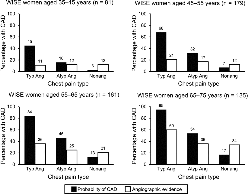
Fig. 7.4
Probability of coronary artery disease compared with observed coronary disease prevalence in symptomatic women aged 35–75 Years. Across all age groups and angina presentations, women in the Women’s Ischemia Syndrome Evaluation (WISE) study tended to have discordance between angiographic evidence of actual disease and their probability of having disease. Atyp ang indicates atypical angina, CAD coronary artery disease, Nonang nonangina, Typ Ang typical angina (Adapted from Shaw et al. [57]. Used with permission from Elsevier Limited)
Women present more often than men with sudden cardiac death as the sentinel event [65]. Women who present with an acute coronary syndrome have a lower incidence of ST-segment elevation myocardial infarction (STEMI) compared with men (27 % vs 37 %) based on the Global Use of Strategies to Open Occluded Arteries in Acute Coronary Syndrome (GUSTO) IIb trial [66]. Women who presented with acute coronary syndromes had higher elevation than men in markers such as high-sensitivity C-reactive protein and brain natriuretic peptide compared with the traditional cardiac markers such as CK-MB or troponin [67]. Also, when women do undergo percutaneous coronary intervention, they are more likely than men to experience a vascular complication [68].
Acute Coronary Syndromes: Management
The major trials assessing an early invasive vs a conservative approach for management of non–STEMI (NSTEMI) in women report conflicting results. The TACTIMS-TIMI 18 (Treat Angina with Aggrastat and Determine Cost of Therapy with an Invasive or Conservative Strategy–Thrombolysis in Myocardial Infarction 18) trials showed that women with an intermediate or high TIMI risk score tend to benefit from an early invasive strategy [69]. However, women with a low TIMI risk score had an increased risk of adverse events. The FRISC II (Fragmin and Fast Revascularization during Instability in Coronary Artery Disease) study showed that women who presented with unstable angina or STEMI were older, with no difference in rate of myocardial infarction or death in those who were in the invasive vs the conservative arm [70]. The latter finding is in contrast to that for men, who had a better outcome with the invasive strategy.
Given the aforementioned data, the ACC/AHA has made specific recommendations for management of unstable angina or NSTEMI in women. Class I recommendations include pharmacologic management with an antiplatelet regimen and risk factor modification. The invasive strategy is reserved for women with high-risk features, whereas the conservative strategy is for women with low-risk features [71].
Summary of ACC/AHA guidelines for unstable angina and NSTEMI (Class I) [71]:
1.
Women with unstable angina/NSTEMI should be managed with the same pharmacologic therapy as men, both in the hospital and for secondary prevention, with attention to antiplatelet and anticoagulant doses based on weight and renal function; doses of renally cleared medications should be based on estimated creatinine clearance (Level of Evidence B).
2.
Recommended indications for noninvasive testing in women with unstable angina/NSTEMI are similar to those for men (Level of Evidence B).
3.
For women with high-risk features, recommendations for invasive strategy are similar to those for men (Level of Evidence B).
4.
In women with low-risk features, a conservative strategy is recommended (Level of Evidence B).
ST-Segment Elevation Myocardial Infarction
Currently the ACC/AHA guidelines recommend that female patients who present with STEMI be treated similarly to male patients with STEMI [72]. Hasdai et al. [73] showed that cardiogenic shock is more likely to develop in women than in men who present with STEMI. In centers where percutaneous coronary intervention is not available, fibrinolysis should be implemented; however, women who have received fibrinolytics have generally been older, with many more comorbid conditions than men [74, 75]. Thus, women demonstrate a higher mortality and morbidity when treated with fibrinolytics, compared with men, and a higher incidence of bleeding and hemorrhagic stroke. When women undergo percutaneous coronary intervention for STEMI, they seem to have a higher rate of in-hospital mortality, which is most likely due to advanced age [76, 77].
Acute Coronary Syndrome in Women: Treatment and Prevention
Aspirin
All women with significant cardiac risk factors should receive either aspirin for secondary prevention if there are no bleeding contraindications or clopidogrel if there is a contraindication or allergy to aspirin. The Antiplatelet Trialists’ Collaboration reviewed several studies with a combined patient population of 100,000 patients and found that aspirin at a dose of 75–162 mg was beneficial for secondary prevention in women [78].
The data on aspirin use for primary prevention in women are conflicting. The ACC/AHA recommends aspirin 75162 mg daily in high-risk women. The Women’s Health Study was an important study in determining –the benefit of aspirin for primary prevention in women [79]. It randomized women older than age 45 years to 100 mg of aspirin every other day vs placebo and followed them for 10 years. This study showed no difference in the rate of myocardial infarction, but did show a significant reduction in the risk for ischemic stroke. However, there was also an increase in hemorrhagic stroke and gastrointestinal bleeding. The benefit of aspirin in reducing the rate of myocardial infarction was evident only in women older than 65 years of age. The ACC/AHA strongly advises against the use of aspirin in low-risk women [18].
Glycoprotein IIb/IIIa Inhibitors
There appears to be a differential effect of treatment between men and women treated with glycoprotein IIb/IIIa (GPIIb/IIIa) inhibitors. The PURSUIT (Platelet Glycoprotein IIb/IIIa in Unstable Angina: Receptor Suppression Using Integrilin Therapy) trial showed that women had an increased rate of myocardial infarction or death at 30 days compared with men [80]. In the CRUSADE (Can Rapid Risk Stratification of Unstable Patients Suppress Adverse Outcomes with Early Implementation of the ACC/AHA Guidelines) registry, similar results were seen; however, this was mostly due to the use of higher doses of medications that resulted in more bleeding, which is more common in women [81]. Women were perhaps more likely to have received too high a dose, without consideration of such factors as body size, renal function, and advanced age. Thus, females who present with acute coronary syndrome with negative biomarkers would not benefit from GPIIb/IIIa and might even have an increased risk of bleeding, myocardial infarction, or death unless other high-risk features are present that would mandate dose adjustment of GPIIb/IIIa agents.
Stay updated, free articles. Join our Telegram channel

Full access? Get Clinical Tree


