Fig. 9.1
(a) Rotated contrast-enhanced 3D MR angiogram of cervical arteries. The arrowheads indicate the right VA. (b) Longitudinal plane of the VA (V0 through V3 segments) with superimposed color flow. Note the acoustic shadow of the transverse process between the V2 segments (small arrow). (c) Doppler spectra depict characteristic waveforms for each VA segment. Note the higher resistance and pulsatility in the VA origin due to proximity to the subclavian artery (SA) as opposed to the low-resistance waveforms in more distal VA segments
Extracranial Duplex Ultrasound Examination Technique and Scanning Protocol
The extracranial duplex examination allows assessment of the VA mid-cervical portion (V2), the proximal VA and its origin (V0/V1), and the most distal portion accessible on the neck (V3). A linear transducer with 4–7-MHz frequency range is commonly used. To optimize the gray scale image on B-mode, set the dynamic range to 40–50 dB, and the time-gain compensation (TGC) as appropriate to the depth of the vertebral arteries. The B-mode image helps with the assessment of the diameter of the VA (which is usually about 3–4 mm). It may also detect atherosclerotic plaque, intraluminal clots, or even a double lumen in case of an arterial dissection. However, these findings are less prevalent than in patients with carotid artery disease.
Imaging in the Longitudinal Plane
Patients should be placed in a supine position with the neck extended. To find the VA, first visualize the common carotid artery in a longitudinal projection on B-mode with transducer position anterior to the sternocleidomastoid muscle. Slide the probe posteriorly to image one or more levels of the V2 segment of the VA as it courses through the transverse processes of the vertebrae (“acoustic shadows”) (Fig. 9.1). Confirm that the direction of flow in the VA is the same as the common carotid artery. Then trace the V2 segment proximally and “heel-toe” the probe above the clavicle to image the pre-vertebrae portion and the origin of the VA as it arises from the SA. It is important to examine this part as V0/V1 is the most common site of atherosclerotic disease [21]. Finally, follow the V2 segment of the VA more distally trying to visualize the V3 segment as it surrounds the axis. However, direct assessment of the V3 segment may be technically difficult, and the diagnosis of vertebral obstruction at this level is based on color-flow and Doppler findings proximal or distal to this segment (Figs. 9.1 and 9.2). If color or power flow images are difficult to obtain, spectral interrogation of areas with typical B-mode appearance of surrounding bony structures should still be performed.


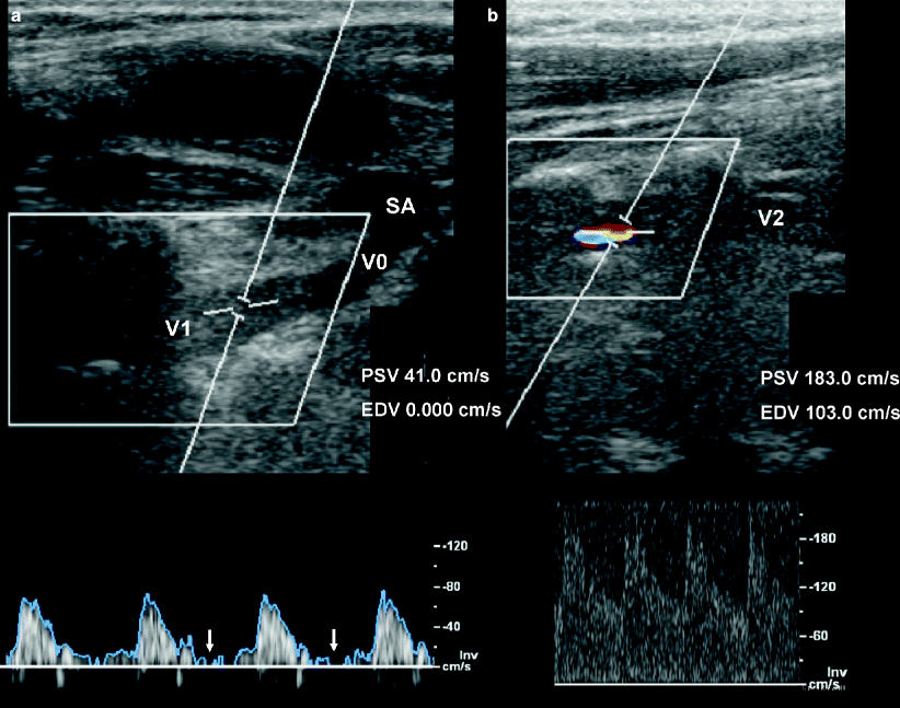
Fig. 9.2
Two case examples. (a) A high-resistance flow in the proximal VA indicates a severe obstruction distally in a patient with an atherosclerotic VA occlusion. Note that there is no end-diastolic flow on Doppler spectra (arrows). (b) Increased focal flow velocity with a severe stenosis due to angiographically confirmed arterial dissection of the mid-cervical VA at C4 and C5 level. Note aliasing artifact on color-flow image
Color-Flow Ultrasound Evaluation of Flow Dynamics
Choose the appropriate color pulse repetition frequency (PRF) by setting the color velocity scale for the expected velocities in the vessel. For normal adult arteries, the peak systolic velocity range is usually between 40 and 50 cm/s. The zero baseline of the color bar is set at approximately two-thirds of the range with the majority of frequencies allowed in the red direction (for flow toward the brain). Adjust the scale further to avoid systolic aliasing (low PRF) or diastolic flow gaps (high PRF or filtering) in normal vessels.
Initially the color or power mode gain should be adjusted to an optimized level, with color displayed throughout the lumen of the vessel and no “bleeding” of color into the surrounding tissue. This is the level at which all color images should be assessed. In situations where there is very low flow, or questionable occlusion, an “over-gained” level may be advantageous to show any flow that might be present, e.g., total occlusion versus a near-occlusion or critical stenosis.
The size of the color box should cover the entire vessel diameter and at least 1–2 cm of its length. Large or wide color boxes will slow down frame rates and resolution of the imaging system. Align the box, i.e., select an appropriate color-flow angle correction, according to the vessel geometry and course.
Doppler Spectral Evaluation of Flow Dynamics
Display the longitudinal image of the VA. Use color-flow image as a guide for Doppler examination. Begin the examination using a Doppler sample volume size of 1.5 mm positioned in the middle of the vessel. The insonation angle should be parallel to the color blood flow and lower than or equal to 60° in every segment. Adjust the Doppler spectral power and gain to optimize the quality of the signal return. Slowly sweep the sample volume throughout the different inter-vertebrae visualized segments. Identify regions of flow disturbance or absence. Additionally, include Doppler spectral waveforms proximal, within, and distal to all areas where flow abnormalities were observed. Locate the origin or proximal segment of the VA. Record flow patterns paying careful attention to flow direction. Follow accessible cervical segments of the VA. Change angulations of the color box and Doppler sample along with the course of the artery.
Following Data Should be Provided
1.
Highest peak systolic velocity in each VA segment
2.
Highest end-diastolic velocity in each VA segment
3.
Flow direction of each VA segment
4.
Documentation of the Doppler spectral waveform pattern from each VA segment
5.
Views demonstrating the presence and location of abnormalities
Tips to Improve Accuracy
1.
Consistently follow a standardized scanning protocol.
2.
Always perform a complete examination of each VA segment.
3.
Sample velocity signals throughout all arterial segments accessible.
4.
Use multiple scan planes.
5.
Take time to optimize the B-mode, color, and spectral Doppler information.
6.
Videotape or create a digital file of the entire study including sound recordings.
7.
Always use the highest imaging frequencies to achieve higher resolution.
8.
Account for any clinical conditions or medications that might affect velocity.
9.
Integrate data from the right and left VA.
10.
Do not hesitate to admit uncertainty and list all causes for limited examinations.
Clinical Indications and Diagnostic Criteria of Vertebral Artery Ultrasonography
In the following, we describe established clinical indications for VA ultrasonography in routine clinical practice, and specific diagnostic criteria derived from previous studies and our own experience (Table 9.1).
Table 9.1
Diagnostic criteria for VA ultrasonography
Diagnostic criteria | B-mode image | Color-flow image | Doppler spectral display |
|---|---|---|---|
≥50% stenosis | Structural lesions (e.g., vessel wall thickening/plaque) when able to directly visualize | Diastolic flow signal void proximal to the lesion | Focal significant PSV increase (usually ≥100 cm/s) with PSR ≥2 |
Flow lumen narrowing when able to visualize | Bruit (turbulences), spectral narrowing (when traceable) | ||
Aliasing artifacts (with proper PRF settings) | Indirect pre- and post-stenotic signs (abnormal pulsatility/waveforms) | ||
Occlusion | Hypoechoic vessel lumen (acute/subacute occlusion) | Flow signal void at occlusion site | Absent or minimal (including systolic/dicrotic notch small spikes) Doppler signals at occlusion site |
Hyperechoic vessel lumen (chronic occlusion) | Diastolic flow signal void proximal to the lesion | Indirect pre- and post-stenotic signs (abnormal pulsatility/waveforms) | |
Segmental occlusion | Hypoechoic vessel lumen (acute/subacute occlusion) | Flow signal void at occlusion site | Antegrade low-resistance flow pre- and post-lesion |
Hyperechoic vessel lumen (chronic occlusion) | Diastolic flow void gap proximal to the lesion | Delayed systolic upstroke distal to the lesion | |
Patent distal VA segment | |||
Nondominant VA | Decreased vessel diameter | Undisturbed flow signals corresponding to a relatively diminished lumen | Velocity lower than contralateral side by 20% or morea |
Normal pulsatility (PI 0.6–1.1)a | |||
Hypoplastic VA | Decreased vessel diameter relative to the other side. | Undisturbed flow signals corresponding to a relatively diminished lumen | Low velocity (PSV < 40 cm/s)a |
High pulsatility (PI ≥ 1.2)a | |||
Arterial dissection | Vessel wall irregularities (when directly visualized) | Flow signal void (with complete obstruction) | Absent or minimal (including systolic/dicrotic notch small spikes) Doppler signal at occlusion site
Stay updated, free articles. Join our Telegram channel
Full access? Get Clinical Tree
 Get Clinical Tree app for offline access

|