Figure 7.2 illustrates the two types of altered or “aberrant” conduction from the atria (PR interval) to the ventricles (QRS interval) that results from bundle-branch block (BBB) and ventricular preexcitation. Right or left BBB does not alter the PR interval, but prolongs the QRS complex by delaying activation of one of the ventricles (see Fig. 7.2A). Ventricular preexcitation, due to a connection of the ventricle to the atria via an accessory muscle bundle, shortens the PR interval and produces a “delta wave” in the initial part of the QRS complex (see Fig. 7.2B). The total time from the beginning of the P wave to the end of the QRS complex remains the same as in the normal condition, because conduction via the abnormal pathway does not interfere with conduction via the normal AV conduction system. Therefore, before the entire ventricular myocardium can be activated by progression of the preexcitation wavefront, electrical impulses from the normal conduction system arrive to activate the remainder of the ventricular myocardium.

The combination of the following has been termed the WPW syndrome.
1. PR interval duration of <0.12 second.
2. A delta wave at the beginning of the QRS complex.
3. A rapid, regular tachyarrhythmia.
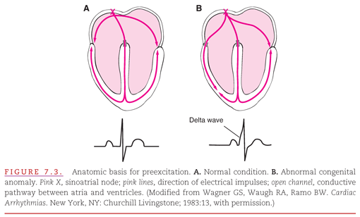
The PR interval is short because the electrical impulse bypasses the normal AV nodal conduction delay. The delta wave is produced by slow intramyocardial conduction that results when the impulse, instead of being delivered to the ventricular myocardium via the normal conduction system, is delivered directly into the ventricular myocardium via an abnormal or “anomalous” muscle bundle. The duration of the QRS complex is prolonged because it begins “too early,” in contrast with the situations presented in Chapters 5 and 6, in which the duration of the QRS complex is prolonged because it ends too late. The ventricles are activated successively rather than simultaneously; the preexcited ventricle is activated via the bundle of Kent, and the other ventricle is then activated via the normal AV node and His–Purkinje system (Fig. 7.3).

The relationship between an anatomic bundle of Kent and physiologic preexcitation of the ventricular myocardium, and the typical ECG changes of ventricular preexcitation, are illustrated on top and on bottom, respectively (see Fig. 7.3). Figure 7.3A illustrates the normal cardiac anatomy that permits AV conduction only via the AV node (the open channel at the crest of the interventricular septum). Thus, there is normally a delay in the activation of the ventricular myocardium (PR segment), as noted in the ECG recording shown in the figure. When the congenital abnormality responsible for the WPW syndrome is present, the ventricular myocardium is activated from two sources via: (a) the preexcitation pathway (the open channel between the right atrium and right ventricle shown in Fig. 7.3B) and (b) the normal AV conduction pathway. The resultant abnormal QRS complex (termed a fusion beat) is composed of the abnormal preexcitation wave and normal mid- and terminal-QRS waveforms.
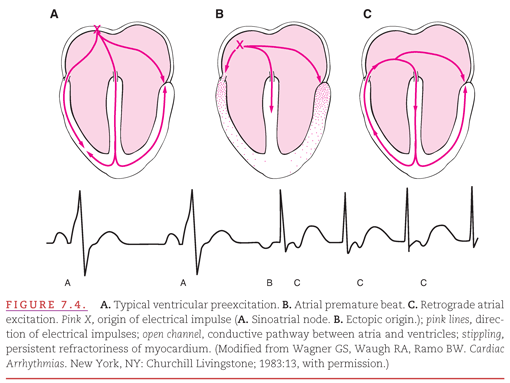
The abnormal AV muscular connection completes a circuit by providing a pathway for electrical reactivation of the atria from the ventricles. This circuit provides a continuous loop for the electrical activating current, which may result in a single premature beat or a prolonged, regular, rapid atrial and ventricular rate called a tachyarrhythmia (Fig. 7.4). In Figure 7.4B, an atrial premature beat has occurred and sends a wave of depolarization through the atria and toward the bundle of Kent. Because this beat originated in such close proximity to the bundle of Kent, the bundle has not had sufficient time to repolarize. As a result, the premature wave of depolarization cannot continue through this accessory AV conduction pathway to preexcite the ventricles. However, the premature wave is able to progress to the ventricles via the normal AV conduction pathway in the AV node and interventricular septum. This depolarization wave then travels through the ventricles, and because it does not collide with an opposing wave (as occurs with ventricular preexcitation in Fig. 7.4A), it reenters the atrium through the bundle of Kent, creating a retrograde atrial excitation (see Fig. 7.4C).

Ventricular preexcitation, induced by an accessory pathway, influences the ventricular rate to become rapid in the presence of an atrial tachyarrhythmia such as atrial flutter/fibrillation (see Chapter 17
Stay updated, free articles. Join our Telegram channel

Full access? Get Clinical Tree


