Ankle-brachial index
≥0.9–1.3
Normal range
≥0.6–0.8
Borderline perfusion
≤0.5
Severe ischemia
Ankle pressure ≤40 mmHg
Critical limb ischemia
Toe pressure ≤30 mmHg
Critical limb ischemia
Transcutaneous oxygen pressure measurement (TcPO 2 )
>40 mmHg
Sufficient to support ulcer healing
20–40 mmHg
Equivocal wound ulcer healing
<20 mmHg
Inability to support ulcer healing
Pulse volume recordings or Doppler waveforms
Triphasic
Normal
Biphasic
Mild to moderate arterial obstruction
Monophasic
Severe arterial obstruction
24.2.7 X-Ray Evaluation
Evaluation with x-ray helps determine if there is underlying pathology contributing to the severity or cause of the leg ulcer. Patients with clear infection of the foot should undergo plain x-rays of the foot to screen for changes in the bone consistent with osteomyelitis. Computed tomography or magnetic resonance imaging can help determine the presence of deep infection, the presence of soft tissue mass, or the presence of an arteriovenous malformation. Radionucleotide scans can sometimes be helpful in also determining the presence of occult or deeper infected tissue or abscess.
24.3 Venous Ulcers
24.3.1 Epidemiology
Venous ulcers are the most common cause of leg ulcers, representing 45–90 % of all leg ulcers [6–9]. Although the exact prevalence of venous ulcers is unknown, the range is estimated to be between 1 and 3 % of the general population in developed countries [11–14]. Of the six to seven million people in the United States with chronic venous disease, about one million individuals have venous ulcers. Contracting these ulcers leads to major economic implications with the lifetime average cost exceeding $40,000 and total cost of care in the United States exceeding one billion dollars per year [7, 14, 15]. These costs are further exacerbated at all levels due to the high recurrence rates of venous ulcers which can be as high as 57–97 % [12].
24.3.2 Pathophysiology
Venous ulcers are a direct result of venous ambulatory hypertension from chronic venous disease. The causes of chronic venous disease include venous insufficiency from valve reflux, venous obstruction, calf muscle pump dysfunction, or a combination of two or more of these causes. The end result is venous hypertension in the upright position which leads to congestion of the deep and superficial venous system. This increase in pressure is ultimately transmitted to the capillaries which in turn leads to leaking of protein-rich serum and erythrocytes into the subcutaneous space. This leads to migration of neutrophils and a pronounced inflammatory response. The physical signs of this process are edema, dermatitis, hyperpigmentation, lipodermatosclerosis, and ulceration. The large majority of venous ulcers occur in the gaiter area of the leg around the medial or lateral malleolus. These ulcers can occur spontaneously or begin after a minor trauma.
24.3.3 History and Physical Examination
Past history of deep venous thrombosis remains a vital part of determining whether chronic venous disease may play a role in a chronic leg ulcer. Location and when the DVT occurred should be noted. History should also include probing questions as to whether the patient may have had an occult deep venous thrombosis. This would include past trauma or surgery to the leg, particularly orthopedic surgery. Historical features unique to deciphering whether venous disease plays a role in a leg ulcer include a past or present history of deep venous thrombosis, pregnancy, obesity, arthritis or other conditions affecting calf muscle pump, varicose veins, trauma to the leg, sedentary lifestyle with chronic dependency of the leg, or a known hypercoagulable state.
Many of the findings on physical examination of leg ulcer are unique only to chronic venous disease. Edema in the early stages can be pitting and then over a longer time becomes “brawny,” or nonpitting. The location of edema in chronic venous disease is in the lower leg below the knee and down to the ankle. Edema from chronic venous disease does not involve the foot. Hyperpigmentation occurs primarily in the ankle area from chronic deposition of erythrocytes that are broken down and deposit hemosiderin [16, 17]. Telangiectasias, reticular veins, and varicose veins are often present. Stasis or venous dermatitis results from the pronounced inflammatory response in the dermis and epidermis. The skin can appear with erythema, scaling, crusting, weeping, and erosions. These areas can incite extreme pruritus, and subsequent scratching can cause mild breaks in the skin which can become a nidus for venous ulceration formation and/or cellulitis. Lipodermatosclerosis can be noted to form in the gaiter area of the leg. This fibrosis and hardening of the subcutaneous space can begin to limit how much edema can occur and subsequently cause the lower leg to appear like an upside-down champagne bottle. These fibrotic changes are thought to be a combination of chronic inflammation and deposition of fibrin and collagen. In some areas within the ankle, smooth white plaques on the skin can occur which are called atrophie blanche. These lesions can be a precursor to venous ulceration.
The venous ulcer is classically located around or on the medial or lateral malleolus (Fig. 24.1). These ulcers can vary in size depending on the chronicity. They are typically exudative and shallow with a dark-red base or possess yellow necrotic slough. The skin borders are generally irregular and maceration and intense skin inflammatory changes are also present. Intensity of pain and tenderness varies from patient to patient.
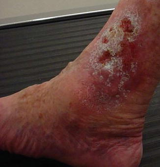
Fig. 24.1
Classical location for a venous ulcer near the medial or lateral malleolus
24.3.4 Management
The mainstay of therapy for the treatment of chronic venous disease and healing venous ulcers is compression therapy [18, 19]. Used as early as the seventeenth century, the application of externally applied pressure or static support helps to return the venous system back to a “normal physiologic state.” There are multiple types of compression therapy and all have certain advantages and disadvantages. The basic mechanism of action provides graduated pressure from the ankle to the knee and augments the dysfunctional calf muscle pump during ambulation. With every dorsiflexion, venous return is augmented. Compression also increases the interstitial pressure in the subcutaneous space and muscular compartments while partially collapsing superficial veins. This action promotes normal function of the venous valves, increases velocity of blood flow, and reduces aggregation and extravasation of neutrophils [20, 21].
Compression devices can be categorized as either sustained/static or intermittent/dynamic. Sustained or static compression includes different types of compression wraps, stockings, or orthoses. Intermittent or dynamic compression includes pneumatic pump devices. Table 24.2 lists the breakdown of types of compression with advantages and considerations. Generally, when a patient presents with a venous ulcer, the leg is edematous, weeping, and macerated. Multilayered compression wraps such as Profore (Smith and Nephew, London, England) with multiple layers of elastic and nonelastic components including a thick layer of gauze are the early choice of treatment. Advantages include the ability to absorb exudate, adapt to a changing leg diameter while maintaining compression, and provide continuous compression either with ambulation or at rest. Once the edema is reduced considerably, skin integrity is improved, and the ulcer has decreased in size, maintenance compression can be started. For elderly individuals, patients with weakened upper extremity strength, or the morbidly obese, an orthosis such as CircAid (Coloplast Corp, Marietta, Georgia) may be the best option to continue ulcer healing while maintaining control of edema. Younger patients may do better with transition to elastic compression stockings for maintenance therapy. In the United States, elastic compression stockings come in four classes, depending on their strength of support (Table 24.3). In the large majority of patients, below-knee compression suffices for treatment of a venous ulcer when in a maintenance stage.
Table 24.2
Classification of types of leg compression that can be used for chronic venous disease and the venous leg ulcer
Sustained (static) | ||
|---|---|---|
Subtypes | Advantages | Considerations |
Elastic | ||
Multilayered compression wraps | For early treatment to control exudates and edema, for sedentary or ambulating patients | Requires skilled personnel to apply |
Single-layer elastic wraps | Early treatment or maintenance therapy | Applied by trained caregiver, reusable |
Compression stockings | Later treatment or maintenance therapy | Needs fitting, requires patient skill and strength to apply |
Inelastic | ||
Unna boot | Early treatment for edema and exudate, good for ambulating patient | Applied by trained caregiver |
Short-stretch wrap | Early treatment or maintenance therapy, good for ambulating patient, permissible for patients with ABIs of 0.5–0.8 | Applied by trained caregiver, reusable |
Orthoses | Maintenance treatment, used when edema is controlled, easier to apply | Requires fitting, bulky |
Intermittent (dynamic) | ||
Pneumatic compression | Applicable to patients with arterial or venous disease | Avoid in patients with heart disease |
Table 24.3
Levels of compression for elastic stockings
Class | Strength (mm Hg) |
|---|---|
I | 20–30 (light) |
II | 30–40 (moderate) |
III | 40–50 (strong) |
IV | 50–60 |
Not every patient should have compression therapy instituted as there are two primary contraindications. Patients with uncompensated heart failure can be put into acute pulmonary edema with mobilization of interstitial fluid into the intravascular space. Patients with ankle-brachial indices less than 0.5 can have further tissue compromise if high-level elastic compression (30–40 mmHg) is applied. Application of this strength of compression stocking can also incite ischemic rest pain. This type of therapy should only be used on patients with an ankle-brachial index greater than 0.8. For patients between 0.5 and 0.8, moderate compression therapy may suffice. It is also important that arterial perfusion be corrected if the ABI is less than 0.8. This will improve healing potential of the venous ulcer as well as allow the use of a higher level of compression therapy.
The medication pentoxifylline has been shown to increase healing of venous ulcers. One meta-analysis of nine prospective trials comparing the drug to placebo showed significantly better healing with a relative risk of 1.30 (p < 0.05) [22]. The mechanism of healing action of this rheologic is thought to be through the inhibition of cytokine-mediated neutrophil activation. Micronized purified flavonoid fraction (MPFF; trade name Daflon, Servier Company, Neuilly-sur-Seine, France) is thought to increase venous tone. A meta-analysis of randomized placebo-controlled trials showed a relative risk reduction in ulcer healing by 32 % with a shorter time to healing of 16 versus 21 weeks (p < 0.05) [23]. Pale sulfonated shale oil (PSSO) applied topically has been shown to enhance proliferation of growth factor expression and stimulate wound healing. A multicenter, randomized, observer-blinded, placebo-controlled study demonstrated a significant reduction in ulcer size in treatment group versus control group [24]. One hundred and nineteen class 6 patients received 20 weeks of treatment. Results showed the treatment arm went from a mean ulcer size of 15–6.2 cm2 and placebo arm from a mean of 11.4–10.8 cm2. While MPFF and PSSO are not available in the United States, patients with venous ulcers should be started on pentoxifylline 400 mg by mouth three times a day. The most common side effect is generalized stomach upset.
The human skin equivalent Apligraf (Organogenesis, Canton, Massachusetts) has been shown in a prospective randomized trial to have greater healing of venous ulcers compared to a placebo arm at 6 months (63 % versus 49 %, p = 0.02) [25]. Additionally, the median time to complete ulcer closure was significantly shorter at 61 days versus 181 days (p = 0.003). Apligraf is manufactured from cultured human fibroblasts and keratinocytes derived from donated foreskin with each sheet derived from a single donor. Apligraf can only be applied to the venous ulcer when bioburden has been dramatically decreased and the ulcer bed is free of necrotic tissue or fibrinous debris. Apligraf is believed to have three mechanisms of possible action. First, the skin graft may “take” with persistence of graft cells in the wound which stimulate increased healing. Second, cells within Apligraf detect a wound and begin to secret growth factors that stimulate healing. Third, the graft serves as a semipermanent bio-occlusive cover to the wound bed [26]. Apligraf is indicated after a venous ulcer has failed to show any remarkable healing after 1 month of conventional (compression) therapy. Apligraf is applied to the wound with the appropriate bolster dressing in addition to conventional multilayer compression bandage. Compression should be changed weekly and more frequently if the wound is exudative. Up to five applications can be used for the treatment of venous ulcer.
Stay updated, free articles. Join our Telegram channel

Full access? Get Clinical Tree


