Fig. 41.1
Reflux in the GSV in the lower thigh is demonstrated with color flow (left panel) and with Doppler waveform (right panel). Red flow (positive signal) going toward the transducer in the GSV is retrograde flow indicating reflux. The duration of the retrograde flow is >4 s as seen by the Doppler waveform
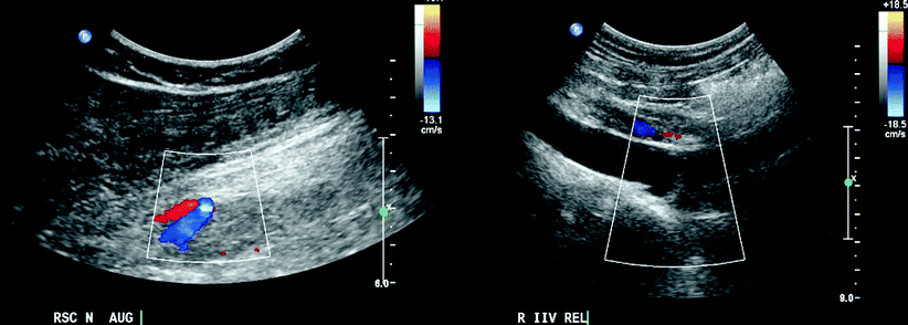
Fig. 41.2
Use of lower frequency transducer to examine deeper veins in thigh and pelvis. Examination of the sciatic nerve veins in the posterolateral thigh is seen on the (left panel). Two veins are seen below the sciatic nerve. The right internal iliac vein is normal during the Valsalva maneuver on the (right panel)
Previous studies utilized diverse methodologies for defining cutoff values regarding Doppler velocities and physiologic reflux time. The vast majority of these studies utilized either the cuff deflation and/or the Valsalva maneuver to establish cutoff values for reflux >500 ms [5–11]. The only study that evaluated most of the lower extremity veins (16 vein sites examined on each limb), with the largest sample size (n = 80 healthy limbs and n = 60 CVD limbs), provided the following cutoff values for venous reflux: reverse flow >500 ms for superficial, deep calf veins, and deep femoral vein >1,000 ms for common femoral, femoral, and popliteal veins (PVs), and >350 ms for perforating veins (Fig. 41.3) [1]. This study demonstrated that patients should be examined in the erect position in order to increase the yield of the DUS examination for the detection of reflux [1].
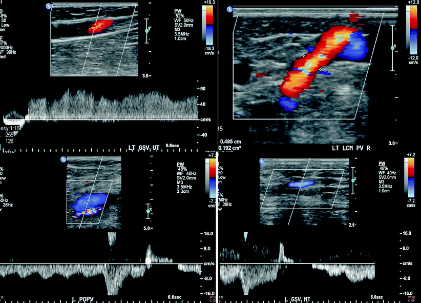

Fig. 41.3
High velocity prolonged reflux is seen in the thigh segment of GSV (upper left panel). A dilated incompetent lower calf perforator measuring 4.9 mm from the same patient is shown on the (right upper panel). Physiologic flow reversal is seen in both the popliteal vein (left lower panel) and the GSV (right lower panel). The duration of retrograde flow was <1 s in the popliteal vein and <0.5 s in the GSV as shown by the timescale (distance between two large divisions is 1 s)
DUS may further contribute in the classification of chronic venous disease into primary, secondary, or congenital based upon the pathophysiology of the detected reflux. Primary chronic venous disease, without a demonstrable cause, is by far the most common type (64–79%) compared to the secondary (18–28%) that is frequently the sequelae of deep venous thrombosis (DVT). Congenital reflux (<5%) is rarely recognizable early on, although it exists since birth, due to an insulate in early symptoms and signs [2, 12].
General Ultrasound Examination and Clinical Considerations
Evaluation of venous reflux should start with the patient in the standing position, facing toward the examiner, and with the examined limb being in slight flexion and external rotation. If the patient is unable to stand, the veins from the midthigh and below can be assessed in the sitting position. If the test is performed on a bed, the torso should be elevated >45°. The supine position is inappropriate for detection of reflux and measurement of vein diameters [1, 12]. Compression with release distal to the point of examination is referred to as augmentation. The former may be applied with several methods: release after a calf squeeze for proximal veins or a foot squeeze for calf veins, manual compression of vein clusters, pneumatic cuff deflation, active foot dorsiflexion and relaxation, and Valsalva maneuver. The vein behaves as a low pressure, collapsible conduit: with compression, its flow is increased in a distal-to-proximal direction and upon release, the blood flow reverses instantaneously [2]. Hence, if valves are competent no retrograde flow is documented. In contrast, if valves are incompetent blood continues to flow in the opposite direction. In an effort to standardize procedures, automated pneumatic cuffs with rapid inflation and subsequent rapid deflation are used [12]. However, in patients with increased BMI or significant edema, the compression technique is difficult to apply, therefore dorsiflexion/plantar flexion or the Valsalva maneuver can be used. The latter is commonly used to evaluate the groin veins especially if the initial compression technique applied provides negative results.
DUS examination protocol starts with the CFV, femoral with deep femoral veins, and continues with the saphenofemoral junction (SFJ) at the terminal and preterminal valves and the associated tributaries, followed by the popliteal vein (PV) and deep calf veins. Thereafter, the superficial veins examined include: great saphenous vein (GSV), small saphenous vein (SSV), their tributaries, and the nonsaphenous veins (Fig. 41.4). Two layers of fascia surround the GSV and with ultrasound imaging (transverse) is referred to as the saphenous eye. SSV is located in the triangular fascia, surrounded by the crural fascia and the gastrocnemius muscle heads. Perforator veins of the lower extremity are usually the last to be evaluated by DUS. They connect the superficial and deep veins traversing the deep fascia, which is visualized easily on B-mode due to its echogenic, collagen-derived structure. Outward flow in perforator veins is seen only in conjunction with superficial and deep vein reflux.
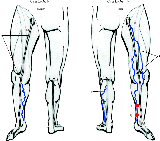

Fig. 41.4
Duplex ultrasound report showing the distribution and extent of reflux. The patient presented with varicose veins in both extremities but she was symptomatic only in the left side. She had burning sensation, itching, and pain on prolong standing that was relieved with limb elevation. On the right side, reflux was found in the GSV from the upper thigh to the knee and at a medial calf tributary of the GSV. SSV and deep veins were normal. On the left side, there was reflux throughout the extent of GSV, in the anterior accessory saphenous vein, a medial calf tributary, two medial calf perforator veins, SSV throughout its length, and a lateral calf tributary. The deep veins were normal
Anatomic variations in both the superficial and deep veins are common. Careful examination and identification of these variations is necessary. Duplication of the PV and femoral vein is common and rare in GSV and SSV. Triple systems may be seen (popliteal, and femoral), and some veins may be hypoplastic or aplastic (e.g., posterior tibial, GSV, and SSV) [13, 14].
Reflux confined to the superficial veins often has a mild to moderate clinical presentation (C1–C3), while involvement of the deep and perforator veins is more often associated with skin damage (C4–C6) [15]. Patients with chronic venous disease exhibit 80% reflux, 17% reflux and obstruction, and only 2% obstruction alone [16]. Old thrombus and/or scar tissue often produces bright echoes within the lumen of the vein of similar brightness to surrounding tissues. In cases of partial recanalization, flow channels are seen and are often incompetent. The vein lumen may be reduced in size and is sometimes not well seen by DUS throughout its length. The vein walls may be thickened and collateral veins are often seen around the area of obstruction (Fig. 41.5) [2].
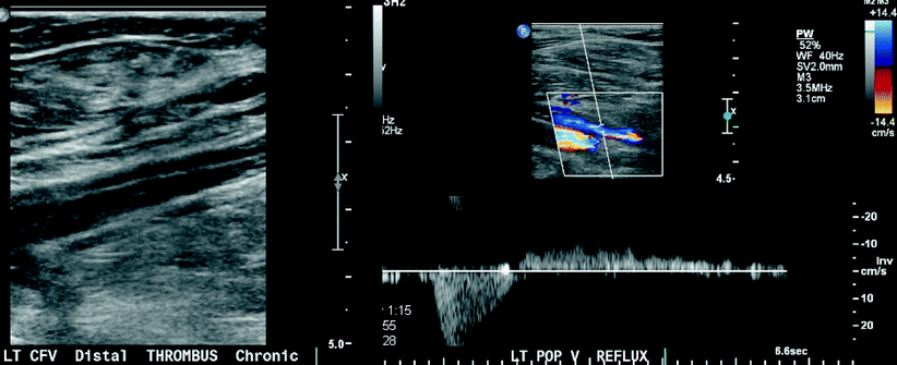

Fig. 41.5
Post-thrombotic changes in the left common femoral vein (left panel). Intraluminal echoes are seen demonstrating partial recanalization. Popliteal vein reflux on the same patient (right panel). The vein is partially recanalized and has prolonged reflux
The combination of obstruction and reflux has a worse prognosis for the development of skin lesions, also known as lipodermatosclerosis [17–20].
Recently, ultrasound-guided-catheter-directed thrombolysis, which involves administration of thrombolytics directly through the side ports of a catheter traversing the thrombus, has been employed in the treatment of venous thrombosis. This technique has been shown to reduce the incidence of post-thrombotic syndrome. A randomized trial has compared catheter-directed thrombolysis followed by 6 months of warfarin with medical treatment alone (intravenous heparin followed by warfarin). This study enrolled 35 of 207 screened patients with acute iliofemoral DVT. Six months after treatment, the patency rate was significantly higher in the group that received catheter-directed thrombolysis (72% vs. 12%), and the prevalence of venous reflux was significantly lower (11% vs. 41%) [21]. Furthermore, the largest observational series from the National Deep Venous Thrombosis Registry demonstrated that patients with iliofemoral DVT had significantly greater 1-year patency rates than patients with femoropopliteal DVT (64% vs. 47%) [22]. The above data demonstrate the multifactorial role of DUS in the diagnosis and monitoring of venous disease.
Site-Specific Ultrasound Examination
Great Saphenous and Accessory Saphenous Veins
The standard ultrasound examination in the groin should begin by identifying the saphenofemoral junction (SFJ), which is located medially to the common femoral artery. Adjacent to the SFJ, various tributaries can be visualized along with the terminal and preterminal valves of the GSV [23, 24]. Reflux is generated by various sources such as SFJ incompetence or lower extremity and pelvic vein insufficiency. The latter is suspected whenever there is a sudden increase in GSV diameter and represents one of the signs of pelvic congestion syndrome along with reflux, which is defined as chronic pelvic pain caused by incompetent ovarian veins. Approximately 10% of women have incompetent ovarian vein valves and of them around 40% experience chronic pelvic signs and symptoms directly as a result of venous congestion (Fig. 41.6) [25–27]. Also, imaging should include the inguinal lymph node area, as well as the full length of the GSV and associated tributaries should be followed up to the ankle. Documentation of the GSV diameter and its distance from the skin is essential since these are used to guide endovenous treatment options [28].
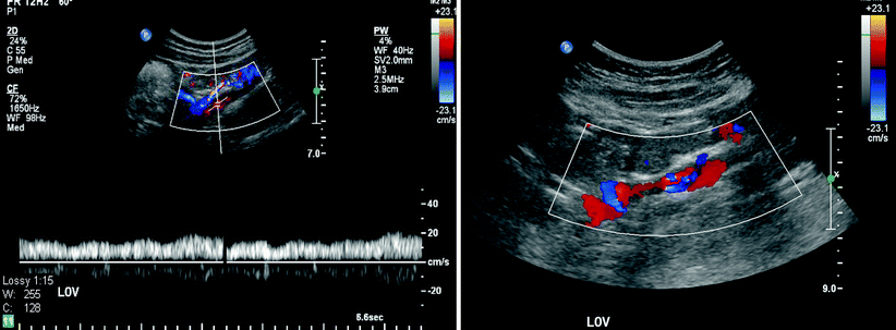

Fig. 41.6
Reflux in the left ovarian vein in a female patient who had two pregnancies and presented with pelvic pain (left panel). Multiple incompetent ovarian veins are seen in the (right panel) in a female patient who presented with pelvic pain and dysuria
Small Saphenous Vein and Its Thigh Extension
Examination starts in the popliteal fossa with the patient standing. Transverse views to visualize the veins within the popliteal fossa and longitudinal views to identify the presence of the saphenopopliteal junction (SPJ) are applied. It is important to rule out SPJ incompetence with SSV reflux [28]. The latter may occur during calf muscle contraction or compression (systolic phase) and suggests possible PV and/or FV obstruction; however, reflux is typically more obvious during calf release (diastolic phase) [29]. The SSV may join the PV medially, posteriorly, or laterally, hence it is advisable to document its location in relation to the PV circumference [30]. A central interconnection element of the local venous circuit is the SSV thigh extension that most often unites GSV near the groin [29]. Documentation of the reflux direction from SFJ to SSV or from saphenopopliteal joint (SPJ)/SSV to GSV should be recorded [31].
Perforator Veins
Veins are identified as perforators only if they pierce through the deep fascia. Their locations are recorded as above or below the knee. The above-knee perforators are further divided as upper, middle, and lower thigh. Those in the below-knee segment are divided as upper, middle, and lower thirds of the calf [32]. Both transverse and oblique scanning are used to evaluate these veins because their long axis is visualized in these planes. Reflux or outward flow in perforator veins is observed in combination with the superficial and deep veins. Bidirectional flow may be seen in some perforator veins, however, only the net outward flow (from deep to superficial) is being evaluated to determine reflux [33]. The examination starts from the medial malleolus, following the cross-sectional image of the posterior arch and GSV upward to the knee region. The anterior arch, the anterior and posteromedial accessory veins, and other thigh tributaries are scanned only if they are varicose [34]. The SSV is imaged from the lateral malleolus until its insertion in the PV. Medial and lateral varicose tributaries of this vein are also scanned. Finally, any area with varicose veins or a vein cluster is scanned for detecting perforators.
Deep Veins: Deep Thigh Veins – Popliteal Vein – Deep Crural Veins
The CFV is examined in the longitudinal view for phasic flow with respiration, cessation of flow with deep inspiration, possible reflux with the Valsalva maneuver, and flow during the compression of the thigh or the calf. Whenever continuous flow is detected in the CFV, the scanning protocol is extended to the inferior vena cava and to the iliac vessels in search of a possible obstruction [35]. Presence of retrograde flow distal to the SFJ corresponds to true deep venous reflux.
PV is examined in its full length, above and below the SPJ, while its anatomic and hemodynamic relationship with the gastrocnemius vein should be clarified. The last part of the examination of the deep veins of the leg involves the depiction of the deep crural veins with the patient usually in the standing or sitting position. Posterior tibial and peroneal veins should be examined in all patients with past and/or present DVT, as especially the latter are the most commonly affected veins of the calf [36]. These veins are imaged well from the medial aspect of the calf. If the peroneal veins are not seen from this window, then imaging is performed from the posterolateral aspect of the calf.
Nonsaphenous Veins
Nonsaphenous veins are superficial venous segments that are not part of the GSV or SSV system. Nonsaphenous venous reflux occurs more commonly in females with previous pregnancies [37]. Nonsaphenous segments that are usually involved are the gluteal, posterolateral thigh perforator, vulvar, lower posterior thigh, popliteal fossa tributaries, knee perforator, and sciatic nerve veins. Patients are examined in the erect position and DUS evaluation is also directed in identifying possible connections with the saphenous and deep veins [38]. The presence of gluteal and/or vulvar varices directs the DUS examination to the pelvic veins [25]. Adjunctive imaging modalities are magnetic resonance venography, CTV or contrast-enhanced venography with selective views of the left renal vein, ovarian, and iliac veins to increase the diagnostic yield and guide therapeutic interventions in cases of pelvic reflux [25–27].
DUS-Guided Management for Combined Deep and Superficial Reflux
Presence of combined superficial and deep venous reflux is common (∼25%) [38]. Patients with combined deep and superficial reflux might improve clinically when the saphenous system is treated [39]. The ESCHAR randomized study showed that in patients with active ulceration, saphenous treatment combined with compression versus compression alone reduced ulcer recurrence risk at 4 years by 25%; however, it did not increase the wound healing rate [40]. The results of saphenous treatment in patients with combined superficial and deep venous reflux are still vague. Despite the fact that CFV reflux can be corrected in most cases, combined femoropopliteal or popliteal vein reflux corrects less often [40]. Deep venous reflux may arise by both primary valve failure and post-thrombotic valve damage, while venous overload is considered another important factor [38].
Finally, there may be patients who exhibit remaining deep venous reflux despite treatment of the superficial reflux. In these patients, specialized treatment options such as valvuloplasty or axillary vein transfer can be applied [32–34]. However, patients with post-thrombotic valves are difficult to treat [32–35].
Stay updated, free articles. Join our Telegram channel

Full access? Get Clinical Tree


