Fig. 40.1
Photoplethysmographic waveforms (PPG). (a) Normal waveforms. (b) Borderline abnormal with partial attenuation of waveforms. (c) Abnormal with complete flattening of the waveforms
Direct Testing with Duplex Ultrasound
Duplex ultrasound has become increasingly accepted as a diagnostic tool in the evaluation of patients suspected of upper extremity arterial disease and venous thrombosis in the vascular community for well over 10 years. With the recent vast improvements in ultrasound equipment, there has been considerable emphasis placed on the expanded role of duplex imaging in the upper extremity. Advances in gray-scale resolution and color Doppler technology permit direct visualization of thrombus, stenosis, collateral vessels, as well as sensitive spectral waveform analysis [11]. These recent advances have greatly reduced, if not eliminated, in most patients, the previous upper extremity ultrasound challenges of insonating the area under the clavicle. With these advancements in duplex imaging, the noninvasive vascular lab has the potential to help aid in the confirmation, treatment, and follow-up care of patients who present with the clinical suspicion of TOS.
Recent studies have been directed toward defining the role of duplex imaging and color Doppler ultrasound with and without maneuvers in diagnosing patients suspected of vascular TOS. In 2001, Wadhwani et al. [15] reported on the effectiveness of duplex imaging with color Doppler ultrasound with maneuvers in five patients clinically suspected of arterial TOS. In 2009, Pacheco et al. published a case study that demonstrated comparable results of duplex ultrasound examination with abduction maneuvers to diagnose venous TOS [5]. Ultimately, both studies found the changes in Doppler waveforms and velocities with and without maneuvers to be an effective method in confirming vascular TOS.
Our Clinical Experience or Experience from Our Institution
As a referral center for TOS at a university medical center, we have seen and evaluated well over 1,000 patients for TOS in the past 6 years. Working closely with the vascular surgery clinical team, our Intersocietal Commission for the Accreditation of Vascular Laboratories (ICAVL) accredited Non-Invasive Vascular Lab has gained extensive experience in the evaluation, diagnosis, treatment and follow-up of this large volume of TOS patients. A total of 312 patients have undergone thoracic outlet syndrome surgical intervention between January 2004 and June 2010. Noninvasive vascular lab studies were performed on these patients as part of the initial evaluation process based on an established algorithm (see Fig. 40.2).
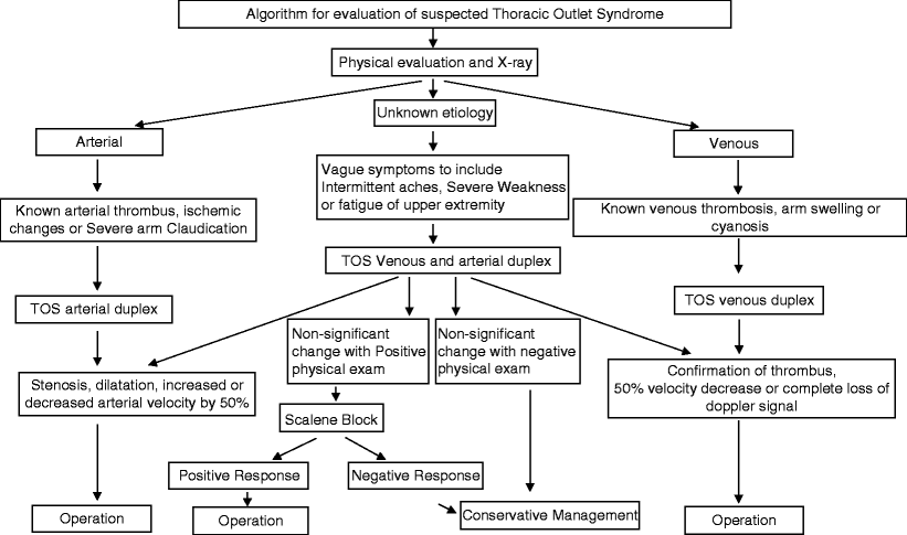
Fig. 40.2
Algorithm for evaluation of suspected thoracic outlet syndrome
TOS Ultrasound Evaluation
All venous and arterial TOS ultrasound examinations were performed by the use of a Phillips iU22 or Phillips 5000 (Bothell, WA) with an 8–4 MHz linear array and 8–5 MHz curved array transducer by a registered vascular sonographer in an ICAVL accredited lab.
TOS Protocol
The examination begins with the patient supine, arms relaxed in the neutral resting position, and without any elevation of the head. Evaluation of the patient’s symptomatic side is performed first with head turned slightly to the contralateral side. The transducer is placed in the transverse plane on the patient’s neck and the internal jugular vein (IJV) is identified. The IJV is examined with gray scale imaging with and without compression cephalad to caudad to evaluate for the presence of occlusive or nonocclusive thrombosis. The IJV is then examined with color Doppler imaging in the longitudinal plane, taking care not to induce manual compression on the vein. Doppler waveform tracings are obtained during quiet respiration. The transducer is then moved back into the transverse plane in the mid-neck region and the IJV is followed inferiorly to the junction of the proximal subclavian vein. The ultrasound probe is placed transversely and slightly medially just superior to the proximal part of the clavicle for the complete evaluation of the proximal subclavian vein. In gray scale, the proximal subclavian vein is examined with and without compression to assess for the presence of occlusive or nonocclusive thrombosis. Doppler angle correction is then utilized in order to obtain waveform and velocity measurements during quite respiration in this proximal region of the subclavian vein. The transducer is then repositioned beneath the clavicle and directed medially to identify and evaluate the mid to distal portion of the subclavian vein as it travels beneath the clavicle in the costoclavicular space [12]. Attention is directed to the location of the subclavian vein as it relates to the subclavian artery. Anatomically, the subclavian vein normally lies superficial and caudal to the subclavian artery and courses with the artery when imaged in the transverse plane [13]. Any deviation in expected anatomic location or inability to identify the subclavian vein should be documented. Identifying the location of subclavian vein to its adjacent subclavian artery is important in differentiating the main vein from enlarged venous collaterals in the subclavicular area (see Fig. 40.3). Gray-scale imaging with and without compression is performed to assess for the presence of occlusive or nonocclusive thrombus in the mid-distal subclavian vein. Color Doppler imaging is then utilized with the probe in the longitudinal plane to evaluate for color filling in the mid through distal subclavian vein. Any reduction or absence of color filling in the vein in this region is documented. Doppler angle correction is then utilized in order to obtain waveform and velocity measurements during quiet respiration in the distal region of the subclavian vein just proximal to the confluence with the cephalic vein.
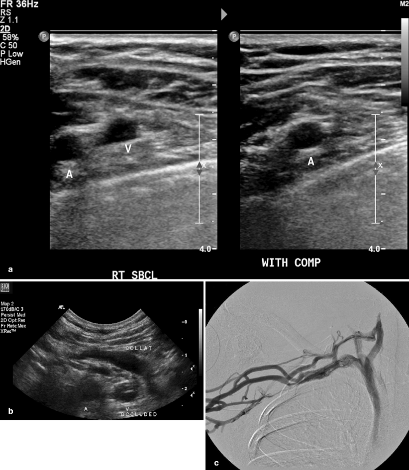
Fig. 40.3
(a) Duplex image of normal anatomic position of subclavian vein and artery. (b) Duplex scan demonstrating abnormal occluded subclavian vein with large collateral vein. (c) Corresponding venogram confirming occluded subclavian vein with large collateral
Next, duplex ultrasound imaging is performed to evaluate the axillary vein. For consistency purposes within a lab, the axillary vein is defined as the vein just distal to the confluence of the cephalic and subclavian vein. The axillary vein continues to the armpit or axilla. The axillary vein lies medial and inferior to the axillary artery in its normal anatomic position. It is important to identify the location of the axillary vein in relationship to the axillary artery in transverse. This view is necessary in order to differentiate the axillary vein from the presence of any possible enlarged venous collaterals. The cephalic vein can be confused with the axillary vein in the clinical setting of axillary vein thrombosis [11]. Both gray scale and color Doppler imaging are performed with and without compression to asses for occlusive or nonocclusive thrombus of the axillary vein. Doppler analysis with angle correction is then utilized in order to obtain waveform and velocity measurements during quite respiration in the axillary vein.
All studies are bilateral using the same TOS venous protocol described previously. We have noted from our experience, similar to the Rochester, NY group, that a significant number of patients experience venous compression of the contralateral side warranting a bilateral examination [14].
For patients who are suspected to have an arterial component of TOS by means of physical and clinical examinations, a scanning technique similar to the venous TOS protocol is employed. As with venous TOS examination, the anatomic location and course of the upper extremity vessels should be carefully noted and documented. Examination in gray scale and color flow imaging of the subclavain artery and axillary artery is done to assess for nonocclusive or occlusive thrombus and evidence of stenosis or vessel dilatation. Angle-corrected Doppler analysis and velocity measurements are documented in the subclavian and axillary artery.
Stay updated, free articles. Join our Telegram channel

Full access? Get Clinical Tree


