Increasing age
Immobility, paralysis
Previous DVT
Malignant disease
Surgery
Trauma
Obesity
Pregnancy
Estrogen therapy
Nephrotic syndrome
Heart failure
Indwelling venous catheters
Venous Duplex Scanning
Prior to the development of ultrasound scanners, contrast venography was considered the “gold standard” for the diagnosis of lower extremity DVT. Currently, venous duplex scanning is considered the gold standard, and contrast venography is reserved for special circumstances such as when an endovascular intervention is planned [2, 11–14]. Other invasive techniques include CT and MR venography, which are indicated in the evaluation of abdominal and pelvic veins or lower extremity vascular malformations. The other noninvasive techniques are described in Chaps. 36and 37(Table 38.2).
Table 38.2
Comparison of diagnostic modalities for lower extremity DVT
Test | Accuracy | Limitations |
|---|---|---|
Venous duplex | Sens: 93–100% | Operator-dependent, experience |
Spec: 97–100% | ||
Contrast venography | Sens: 100% | Up to 15% indeterminate, invasive |
Spec: 100% | ||
IPG | Sens: 73–96% | Inaccurate if nonocclusive thombus |
Spec: 83–95% | ||
CT venography | Sens: 93–100% | Invasive |
MR venography | Sens: 87–100% | Cost |
Spec: 95–100% | ||
Nuclear scintigraphy | Sens: 21–83% | Invasive |
(I125-Fibrinogen) | Spec: 54–97% | Not available in U.S. |
Indications
The symptoms and signs of acute lower extremity DVT are nonspecific. The most common indications to test for a lower extremity DVT include localized pain, swelling, or redness in the calf or thigh, and calf pain with passive dorsiflexion (Homan’s sign) [2, 13, 14]. In order to decrease the probability of a negative test, many physicians obtain a D-dimer level [15]. D-dimer is a fibrin degradation product released in the circulation when fibrinolysis starts on a formed blood clot. The value of D-dimer testing is when it is negative, meaning that it is unlikely that a blood clot is present. A positive D-dimer is present in many conditions, including trauma, pregnancy, inflammatory conditions, and recent surgery [15–18]. The Wells clinical score uses a point system to assess the probability of a DVT (Table 38.3).
Table 38.3
Wells scoring
Clinical characteristic | Point |
|---|---|
Malignancy | 1 |
Paralysis, paresis, cast | 1 |
Recently bedridden for 3 days or more | 1 |
Localized tenderness along course of a deep vein | 1 |
Entire leg swollen | 1 |
Calf swelling, 3 cm or increased circumference | 1 |
Pitting edema in the symptomatic leg | 1 |
Collateral superficial veins on symptomatic leg | 1 |
Alternative diagnosis at least as likely as DVT | −2 |
Instrumentation
Venous duplex scanning of the lower extremity can be performed with several transducers. In a normal adult of average body weight, a 5–10 MHz linear transducer is selected. In a larger or morbidly obese individual, a 3–5 MHz curved transducer will be required to image a deeper plane [9, 11]. In some patients, it may be necessary to use more than one transducer, a lower frequency for the abdomen and groin, and a higher frequency in the calf. In a complete lower extremity venous duplex examination, all three functions are utilized: B-mode, Doppler flow, and color flow.
Technique
Since most ultrasound equipment is portable, venous duplex scanning can be performed in the vascular lab or at the bedside in the Emergency Department or Intensive Care Unit. The patient is positioned supine, and ideally the bed can be tilted for a reverse Trendelenberg or modified Fowler position. When possible, the thigh and calf are slightly externally rotated. To image the abdominal veins, patients should be fasting for 6 h prior to the exam.
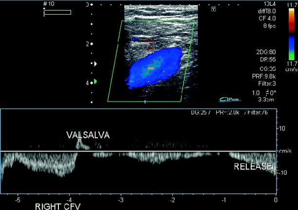
The exam starts by placing the probe in the groin area. Examination of the deep veins, the femoral and common femoral veins, is performed in both the transverse and longitudinal views [9, 11, 14]. The great saphenous vein is imaged as it enters the saphenofemoral junction (Fig. 38.1). With each venous segment identified, gentle compression is exerted to see if the vein walls collapse (Fig. 38.2). This maneuver should be done every 3–5 cm along the length of the medial thigh and popliteal fossa. In addition to compression, Doppler flow is documented by placing the cursor in the middle of the flow channel in longitudinal view. Here, the examiner is observing for spontaneous and phasic flow, as well as the presence of distal augmentation that normally occurs when the calf is gently squeezed (Fig. 38.3). Color-flow is also documented in each segment. For the popliteal vein, the probe is positioned behind the knee and requires that the patient cooperate (Fig. 38.4). All segments of this vein should be thoroughly examined, above, at, and below the knee, with compression, Doppler, and color flow as previously described.
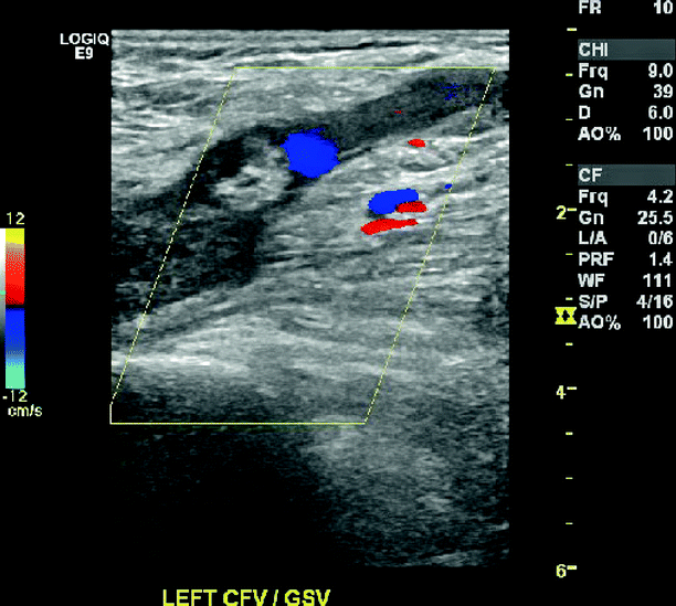
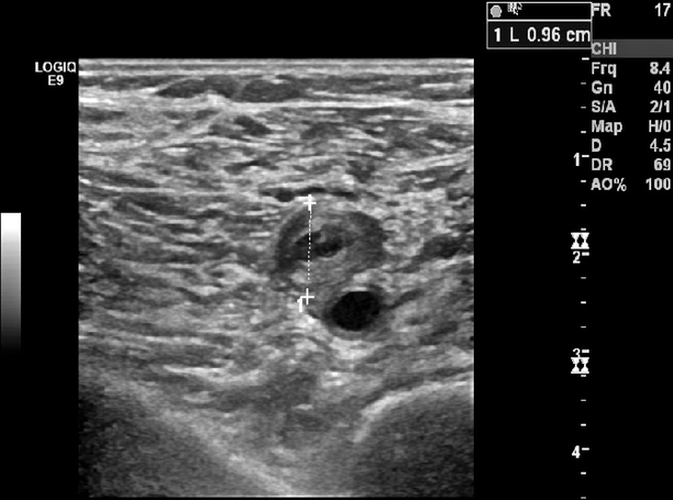
Fig. 38.1
Saphenofemoral junction showing thrombus by B-mode, and near absence of color flow
Fig. 38.2
Acute DVT of the popliteal vein with hyperechoic material filling and distending the vein. The popliteal artery is adjacent showing normal echodensity of flowing blood