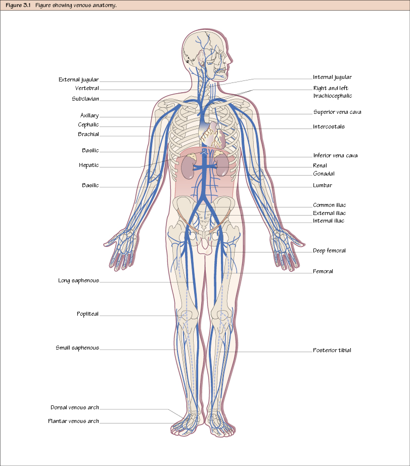
Venous Anatomy

Venous Anatomy

c3-fig-5001

You will need to remember the venous system in the leg because this is the most common site of venous problems (e.g. varicose veins and deep venous thrombosis). However, for vascular access the upper limb and central veins are very important.


Venous Anatomy


The venous circulation is different from the arterial system in the following ways:


  • There is more interperson variability.
  • There is also more functional reserve – we can manage without many of our veins without any ill effect. Even some of the major veins like the inferior vena cava (IVC) can be ligated in an emergency without a devastating effect: blood will find its way back via other routes (i.e. collateral vessels).
  • In keeping with this, there is often more than one vein serving the distribution of one artery, especially in the limbs. These are called venae comitantes and are seen usually as a pair of veins in close relation to an artery and often with many branches between them.
  • In the limbs there is a clear distinction between two sets of veins: the superficial and deep, the former running enveloped by the superficial fascia and the latter running with the arteries.
  • Veins do not have branches, they have tributaries – everything is in reverse order.

Lower Limb


Blood drains from the foot into the dorsal venous arch, which is often visible on the dorsum of the foot. The lateral end of the dorsal venous arch continues as the short saphenous vein and passes posteriorly to the lateral malleolus, lying with the sural nerve. It passes up the posterolateral side of the calf in the subcutaneous fat towards the midline of the leg. It then turns deeply to pierce the deep fascia and continues on to join the popliteal vein at an oblique angle, the join being called the saphenopopliteal junction

Stay updated, free articles. Join our Telegram channel

Jul 1, 2016 | Posted by in CARDIOLOGY | Comments Off on Venous Anatomy

Full access? Get Clinical Tree

Get Clinical Tree app for offline access