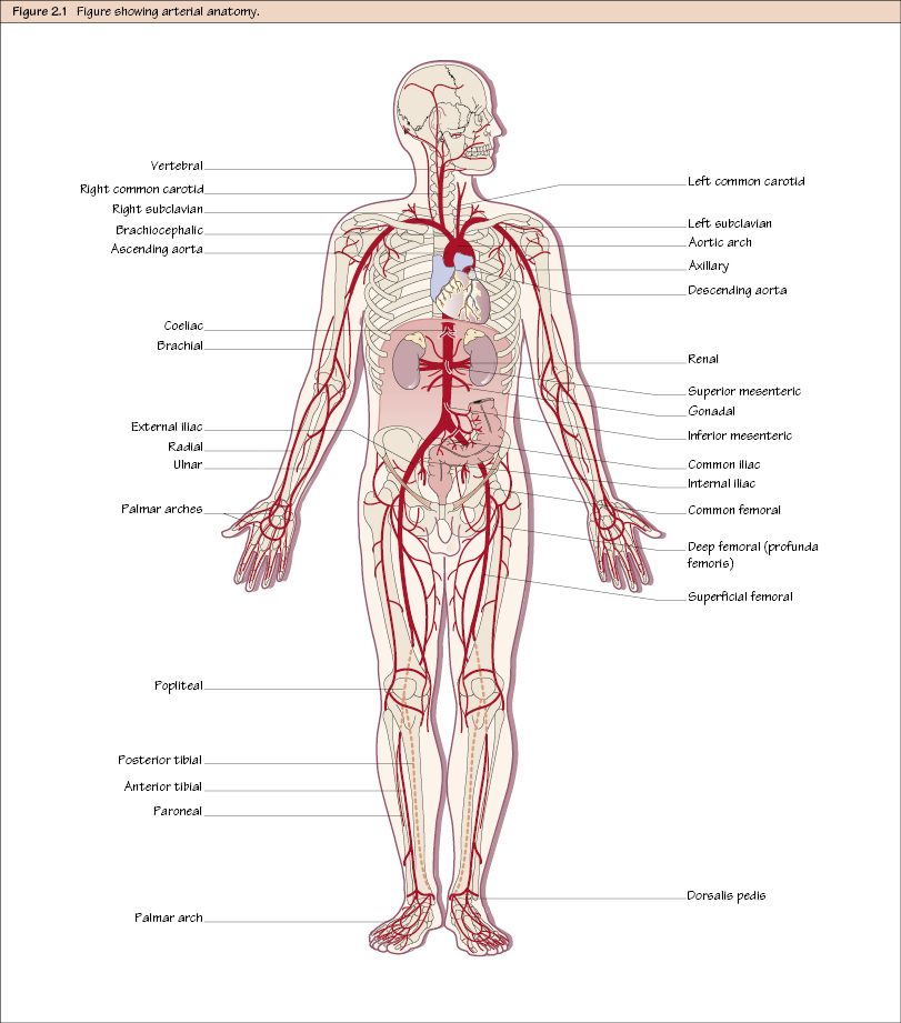
Arterial Anatomy The aorta emerges from the left ventricle at the lower border of the third costal cartilage behind the sternum (slightly to the left). In the superior mediastinum it curves upwards, backwards and to the left, forming in turn the ascending aorta, aortic arch and then the descending thoracic aorta. The ascending aorta gives branches to the heart – the right and left coronary arteries. The outer convexity of the aortic arch gives three branches: On each side then, the common carotid artery ascends in the neck almost and identically passing behind (although very deeply) the sternoclavicular joint to the upper border of the thyroid where it divides into the external carotid and internal carotid. The internal carotid has no branches and ascends into the skull via the carotid canal. The external carotid has several branches supplying the face and neck. Meanwhile, the descending thoracic aorta passes through the thorax on the vertebral column, giving various branches in the mediastinum. It passes through the aortic hiatus in the diaphragm at T12 to become the abdominal aorta. On each side (with the exception of the different origins), the path of the subclavian arteries (SCAs) is basically the same. It travels laterally over the first rib between the anterior and middle scalene muscles, which serve to ‘divide’ it into three different sections, with the second part lying behind the anterior scalene. The branches of the subclavian artery can be memorised by the mnemonic ‘VIT C, D’:
Thoracic and Neck
Upper Limb
Stay updated, free articles. Join our Telegram channel

Full access? Get Clinical Tree


