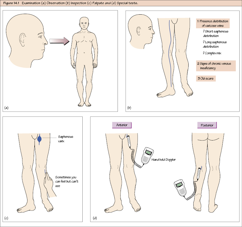
Vascular Examination: Varicose Veins Varicose veins are extremely common, which means you will encounter them in real life and in exams, remarkably frequently. Fortunately examining the venous system is very easy and mainly requires observation and a simple handheld instrument – the Doppler probe. There are a myriad of other ‘special’ tests such as the tourniquet test, which are now considered obsolete because they are totally unreliable. It would be very unfair to ask you to perform them in an exam because they are never used now in clinical practice. The only people who would think it appropriate to include them would be ageing surgical dinosaurs who haven’t practised vascular surgery in decades. Unfortunately, such people still exist in exam contexts, so the tests have been included here in basic format only, just for completeness. The patient should be standing with the knee and hip slightly flexed. The whole leg must be exposed, with only underwear for cover; however, you will need even to look under this for the saphenofemoral junction. You should also be able to see the lower abdomen, as florid varices here suggest IVC obstruction. Inspect the leg looking for:

Observation
Stay updated, free articles. Join our Telegram channel

Full access? Get Clinical Tree


