Vascular Biology There are three basic histological layers (‘tunics’) in a vessel: This is a thin layer consisting of the innermost, single-celled and physiologically active endothelium housed on a dense connective tissue basement membrane (internal elastic lamina). This is the thickest layer of the wall and its content varies according to arterial subtype, anatomical location and exposure to fluid-mechanical stress. It is composed principally of vascular smooth muscle cells (VSMCs) within a connective tissue matrix. This is a poorly defined, heterogeneous, outermost layer of investing connective tissue consisting of a variable amount of smooth muscle cells (SMCs) and fibroblasts along with numerous autonomic nerve endings and vasa vasora (small, microscopic nutritional vessels traversing the layer). Its thickness varies according to location. In large and medium-sized arteries, cells in the innermost media acquire oxygen and nutrition from the blood in the lumen (direct diffusion) while the vasa vasora serve the outer half to two-thirds of the wall. There are two subtypes:
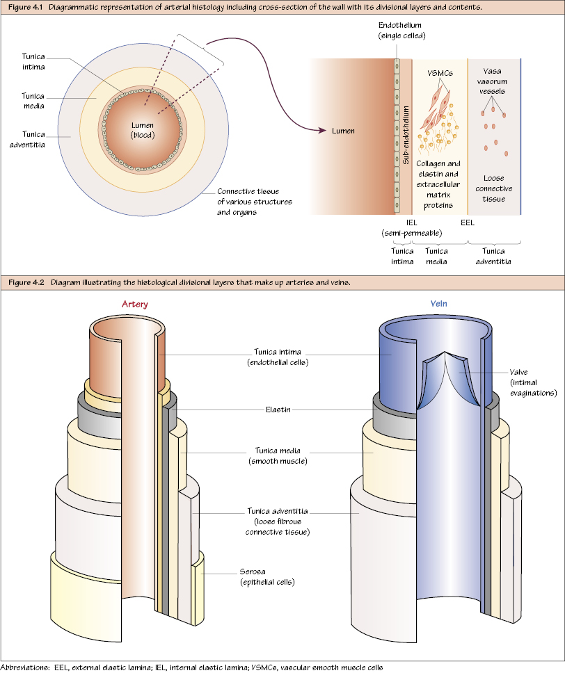
Structure of an Artery
Tunica Intima
Tunica Media
Tunica Adventitia
Blood Vessel Nutrition
Arterial Subtypes
Stay updated, free articles. Join our Telegram channel

Full access? Get Clinical Tree


