Unstable Angina/Non-ST Elevation Myocardial Infarction
Kuang-YuhChyu, MD, PhD Prediman K. Shah, MD
 General Considerations
A. Background
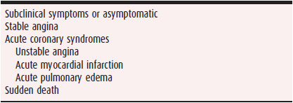
Unstable angina and non-ST elevation myocardial infarction (USA/NSTEMI) are a part of the wide spectrum of clinical manifestations of atherosclerotic coronary artery disease (Table 7–1). Compared with ST elevation myocardial infarction (STEMI), the incidence of USA/NSTEMI has been increasing. According to the National Registry of Myocardial Infarction (NRMI) database, from 1994 to 1999, the prevalence of STEMI decreased from 36.4% to 27.1% with concomitant rise of NSTEMI from 45% to 63%. Similar trends were also observed in a population study from 1999 to 2008. Despite this, the age-adjusted mortality for NSTEMI patients has been gradually declining, likely due to advancement in the treatment for acute coronary syndrome.
Table 7–1. Clinical Spectrum of Atherosclerotic Coronary Artery Disease

B. Pathophysiology
Angina pectoris is the symptomatic equivalent of transient myocardial ischemia, which results from a temporary imbalance in the myocardial oxygen demand and supply. Most episodes of myocardial ischemia are generally believed to result from an absolute reduction in regional myocardial blood flow below basal levels, with the subendocardium carrying a greater burden of flow deficit relative to the epicardium, whether triggered by a primary reduction in coronary blood flow or an increase in oxygen demand. USA/NSTEMI shares a more or less common pathophysiologic substrate with STEMI, which is usually due to ruptured or unstable atherosclerotic plaques with overlying thrombus formation, leading to reduced coronary blood flow. The differences in clinical presentation result largely from the differences in the magnitude of coronary occlusion, the duration of the occlusion, the modifying influence of local and systemic blood flow, and the adequacy of coronary collaterals.
 Clinical Findings
A. Symptoms
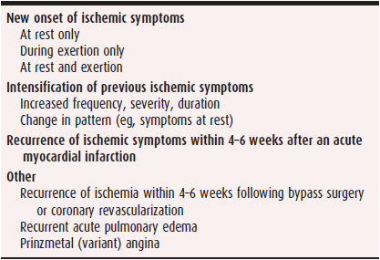
USA/NSTEMI is a clinical syndrome characterized by symptoms of ischemia, which may include classic retrosternal chest pain or such pain surrogates as a burning sensation, feeling of indigestion, or dyspnea (Table 7–2). Anginal symptoms may also be felt primarily or as radiation in the neck, jaw, teeth, arms, back, or epigastrium. The pain of unstable angina typically lasts 15–30 minutes; it can last longer in some patients. In some patients, particularly the elderly, dyspnea, fatigue, diaphoresis, light-headedness, a feeling of indigestion and the desire to burp or defecate, or nausea and emesis may accompany other symptoms or may be the only symptoms. It has been estimated that about 43.6% of patients with USA/NSTEMI presented without chest pain. The clinical presentation of unstable angina can take any one of several forms.
Table 7–2. Clinical Presentation of Unstable Angina

There may be an onset of ischemic symptoms in a patient who had been previously free of angina, with or without a history of coronary artery disease. If symptoms are effort-induced, they are often rapidly progressive, with more frequent, easily provoked, and prolonged episodes. Rest pain may follow a period of crescendo effort angina or may exist from the beginning.
Symptoms may intensify or change in a patient with antecedent angina. Pain may be provoked by less effort and be more frequent and prolonged than before. The response to nitrates may decrease and their consumption increase. The appearance of new pain at rest or with minimal exertion is particularly ominous. On the other hand, recurrent long-standing ischemic symptoms at rest do not necessarily constitute an acute ischemic syndrome. Ischemic symptoms may recur shortly after (usually within 4 weeks) an acute myocardial infarction (MI), coronary artery bypass surgery, or catheter-based coronary artery intervention. In some patients, an acute unstable coronary syndrome may manifest as acute pulmonary edema or sudden cardiac death.
B. Physical Examination
No physical finding is specific for USA/NSTEMI, and when the patient is free of pain, the examination may be entirely normal. During episodes of ischemia, a dyskinetic left ventricular apical impulse, a third or fourth heart sound, or a transient murmur of ischemic mitral regurgitation may be detected. Similarly, during episodes of prolonged or severe ischemia, there may be transient evidence of left ventricular failure, such as pulmonary congestion or edema, diaphoresis, or hypotension. Arrhythmias and conduction disturbances may occur during episodes of myocardial ischemia.
The findings from physical examination, especially as they relate to signs of heart failure, provide important prognostic information. An analysis of data from four randomized clinical trials (GUSTO IIb, PURSUIT, and PARAGON A and B) revealed that Killip classification, a commonly used classification based on physical examination of patients at presentation of STEMI, is a strong independent predictor for short- and long-term mortality, with higher Killip class associated with higher mortality rate. This has also been recently confirmed using data from the Canadian Acute Coronary Syndrome Registries.
C. Diagnostic Studies
USA/NSTEMI is a common reason for admission to the hospital, and the diagnosis, in general, rests entirely on clinical grounds. In a patient with typical effort-induced chest discomfort that is new or rapidly progressive, the diagnosis is relatively straightforward, particularly (but not necessarily) when there are associated electrocardiogram (ECG) changes. Often, however, the symptoms are less clear-cut. The pain may be atypical in terms of its location, radiation, character, and intensity, or the patient may have had a single, prolonged episode of pain, which may or may not have resolved by the time of presentation. The physician should strongly suspect unstable angina, particularly when coronary artery disease or its risk factors are present. When in doubt, it is safer to err on the side of caution and consider the diagnosis to be unstable angina until proven otherwise. Even though dynamic ST-T changes on the ECG make the diagnosis more certain, between 5% and 10% of patients with a compelling clinical history (especially middle-aged women) have no critical coronary stenosis on coronary angiography. In rare instances, especially in women, spontaneous coronary artery dissection, unrelated to coronary atherosclerosis, may be the basis for an acute coronary syndrome. In general, the more profound the ECG changes, the greater the likelihood of an ischemic origin for the pain and the worse the prognosis.
1. ECG and Holter monitoring—ECG abnormalities are common in patients with USA/NSTEMI. In view of the episodic nature of ischemia, however, the changes may not be present if the ECG is recorded during an ischemia-free period or the ischemia involves the myocardial territories (eg, the circumflex coronary artery territory) that are not well represented on the standard 12-lead ECG. Therefore, it is not surprising that 40–50% of patients admitted with a clinical diagnosis of unstable angina have no ECG abnormalities on initial presentation. The ECG abnormalities tend to be in the form of transient ST segment depression or elevation and, less frequently, T-wave inversion, flattening, peaking, or pseudo-normalization (ie, the T wave becomes transiently upright from a baseline state of inversion or vice versa). It must be emphasized, however, that a normal or unremarkable ECG should never be used to disregard the diagnosis of unstable angina in a patient with a compelling clinical history and an appropriate risk-factor profile.
Continuous ambulatory ECG recording reveals a much higher prevalence of transient ST-T wave abnormalities, of which 70–80% are not accompanied by symptoms (silent ischemia). These episodes, which may be associated with transient ventricular dysfunction and reduced myocardial perfusion, are much more prevalent in patients with ST-T changes on their admission tracings (up to 80%) than in persons without such changes. Frequent and severe ECG changes on ECG monitoring, in general, indicate an increased risk of adverse clinical outcome.
2. Angiography—More than 90–95% of patients with a clinical syndrome of unstable angina have angiographically detectable atherosclerotic coronary artery disease of varying severity and extent. The prevalence of single-, two-, and three-vessel disease is roughly equal, especially in patients older than 55 and those with a past history of stable angina. In relatively younger patients and in those with no prior history of stable angina, the frequency of single-vessel disease is relatively higher (50–60%). Left main coronary disease is found in 10–15% of patients with unstable angina. A subset of patients (5–10%) with angiographically normal or near normal coronary arteries may have noncardiac symptoms masquerading as unstable angina, the clinical syndrome X (ischemic symptoms with angiographically normal arteries and possible microvascular dysfunction), or the rare primary vasospastic syndrome of Prinzmetal (variant) angina. It should be recognized, however, that most patients (even those with Prinzmetal angina) tend to have a significant atherosclerotic lesion on which the spasm is superimposed. In general, the extent (number of vessels involved, location of lesions) and severity (the percentage of diameter narrowing, the minimal luminal diameter, or the length of the lesion) of coronary artery disease and the prevalence of collateral circulation, as judged by traditional angiographic criteria, do not differ between patients with unstable angina and those with stable coronary artery disease. The morphologic features of the culprit lesions do tend to differ, however. The culprit lesion in patients with unstable angina tends to be more eccentric and irregular, with overhanging margins and filling defects or lucencies. These findings (on autopsy or in vivo angioscopy) represent a fissured plaque, with or without a superimposed thrombus. Such unstable features in the culprit lesion are detected more frequently when angiography is performed early in the clinical course.
3. Noninvasive tests—Any form of provocative testing (exercise or pharmacologic stress) is clearly contraindicated in the acute phase of the disease because of the inherent risk of provoking a serious complication. Several studies of patients who had been pain-free and clinically stable for more than 3–5 days, however, have shown that such testing, using ECG, scintigraphic, or echocardiographic evaluation, may be safe. Provocative testing is used primarily to stratify patients into low- and high-risk subsets. Aggressive diagnostic and therapeutic interventions can then be selectively applied to the high-risk patients; the low-risk patients are treated more conservatively. In general, these studies have shown that patients who have good exercise duration and ventricular function, without significant inducible ischemia or ECG changes on admission, are at a very low risk and can be managed conservatively. On the other hand, patients with ECG changes on admission, a history of prior MI, evidence of inducible ischemia, and ventricular dysfunction tend to be at a higher risk for adverse cardiac events and therefore in greater need of further and more invasive evaluation.
4. Biomarkers—Blood levels of specific myocardial bio-markers such as cardiac troponins are, by definition, not elevated in unstable angina; if they are elevated without evolution of Q waves, the diagnosis is generally an NSTEMI. This distinction is somewhat arbitrary, however. Patients with negative biomarkers within 6 hours of symptom onset need to have them remeasured within 8–12 hours.
Elevated levels of circulating biomarkers, such as high-sensitivity C-reactive protein, fibrinogen, brain natriuretic peptide, and glucose, have been reported in patients presenting with USA/NSTEMI. The presence of such markers may be useful in risk stratification for clinical outcomes; however, their roles in diagnosing USA/NSTEMI and determining whether treatment strategies based on these biomarkers would alter clinical outcomes have not been established.
Canto AJ, et al. Differences in symptom presentation and hospital mortality according to type of acute myocardial infarction. Am Heart J. 2012;163:572–9. [PMID: 22520522]
Khot UN, et al. Prognostic importance of physical examination for heart failure in non-ST-elevation acute coronary syndromes: the enduring value of Killip classification. JAMA. 2003;290(16): 2174–81. [PMID: 14570953]
Movahed MR, et al. Mortality trends for non-ST-segment elevation myocardial infarction (NSTEMI) in the United States from 1988 to 2004. Clin Cardiol. 2011;34:689–92. [PMID: 22095658]
Rogers WJ, et al. Temporal trends in the treatment of over 1.5 million patients with myocardial infarction in the US from 1990 through 1999: the National Registry of Myocardial Infarction 1, 2 and 3. J Am Coll Cardiol. 200036(7):2056–63. [PMID: 11127441]
Segev A, et al. Prognostic significance of admission heart failure in patients with non-ST-elevation acute coronary syndromes (from the Canadian Acute Coronary Syndrome Registries). Am J Cardiol. 2006 Aug 15;98(4):470–3. [PMID: 16893699]
Shah PK. Pathophysiology of plaque rupture and the concept of plaque stabilization. Cardiol Clin. 2003;21(3):303–14. [PMID: 14621447]
Yeh RW, et al. Population trends in the incidence and outcomes of acute myocardial infarction. N Engl J Med. 2010;362:2155–65. [PMID: 20558366]
 Differential Diagnosis
Conditions that simulate or masquerade as unstable angina include acute MI, acute aortic dissection, acute pericarditis, pulmonary embolism, esophageal spasm, hiatal hernia, and chest wall pain. Careful attention to the history, risk factors, and objective findings of ischemia (transient ST-T changes and mild elevations of troponins in particular) remain the cornerstones for the diagnosis.
A. Acute Myocardial Infarction
Although MI often produces more prolonged pain, the clinical presentation can be indistinguishable from that of unstable angina. As stated earlier, this distinction should be considered somewhat arbitrary because abnormal myocar-dial technetium-99m pyrophosphate uptake, mild creatine kinase elevations detected on very frequent blood sampling, and increases in troponin T and I levels (released from necrotic myocytes) are observed in some patients with otherwise classic symptoms of unstable angina.
B. Acute Aortic Dissection
The pain of aortic dissection is usually prolonged and severe. It frequently begins in or radiates to the back and tends to be relatively unrelenting and often tearing in nature; transient ST-T changes are rare. An abnormal chest radiograph showing a widened mediastinum, accompanied by asymmetry in arterial pulses and blood pressure, can provide clues to the diagnosis of aortic dissection, which can be verified by bedside echocardiography (transesophageal, with or without transthoracic echocardiography), magnetic resonance imaging (MRI), computed tomography (CT) scanning, or aortography.
C. Acute Pericarditis
Acute pericarditis may be difficult to differentiate from unstable angina. A history of a febrile or respiratory illness suggests the former. The pain of pericarditis is classically pleuritic in nature and worsens with breathing, coughing, deglutition, truncal movement, and supine posture. A pericardial friction rub is diagnostic, but is often evanescent, and frequent auscultation may be needed. Prolonged, diffuse ST elevation that is not accompanied by reciprocal ST depression or myocardial necrosis is typical of pericarditis. Leukocytosis and an elevated erythrocyte sedimentation rate are common in pericarditis but not in unstable angina. Echocardiography may detect pericardial effusion in patients with pericarditis; diffuse ventricular hypokinesis may imply associated myocarditis. Regional dysfunction, especially if transient, is more likely to reflect myocardial ischemia.
D. Acute Pulmonary Embolism
Chest pain in acute pulmonary embolism is also pleuritic in nature and almost always accompanied by dyspnea. Arterial hypoxemia is common, and the ECG may show sinus tachycardia with a rightward axis shift. Precordial ST-T wave abnormalities may simulate patterns of anterior myocardial ischemia or infarction. A high index of suspicion, combined with a noninvasive assessment of pulmonary ventilation-perfusion mismatch, evidence of lower extremity deep venous thrombosis, CT angiography, and possibly pulmonary angiography, is necessary to exclude the diagnosis.
E. Gastrointestinal Causes of Pain
Various gastrointestinal pathologies can mimic unstable angina. These include esophageal spasm, peptic ulcer, hiatal hernia, cholecystitis, and acute pancreatitis. A history compatible with those conditions, the response to specific therapy, and appropriate biochemical tests and imaging procedures should help clarify the situation. It should be noted that these abdominal conditions may produce ECG changes that simulate acute myocardial ischemia.
F. Other Causes of Chest Pain
Many patients present with noncardiac chest pain that mimics unstable angina, and sometimes no specific diagnosis can be reached. The pain may be musculoskeletal, or there may be nonspecific changes on the ECG that increase the diagnostic confusion. In these patients, a definite diagnosis often cannot be reached despite careful clinical observation. When the pain has abated and the patient is stable, a provocative test for myocardial ischemia may help rule out ischemic heart disease. Although coronary angiography may provide evidence of atherosclerotic coronary artery disease, anatomic evidence does not necessarily prove an ischemic cause for the symptoms. In some patients, acute myocarditis may also produce chest pain syndromes simulating unstable angina and acute MI. Recreational drug use (cocaine and methamphetamine) may also produce clinical syndromes of chest pain, sometimes related to drug-induced acute coronary syndrome precipitated by the vasoconstrictor and prothrombotic effects of these drugs.
 Treatment
In treating unstable angina, the initial objective is to stratify patients for their short-term morbidity and mortality risks based on their clinical presentations. After risk stratification, management objectives include eliminating episodes of ischemia and preventing acute MI and death.
A. Initial Management
During this early in-hospital phase, therapy is primarily aimed at stabilizing the patient by stabilizing the culprit coronary lesion and thus preventing a recurrence of myocar-dial ischemia at rest and progression to MI.
1. General measures—Patients whose history is compatible with a diagnosis of unstable angina should be promptly hospitalized in an intensive or intermediate care unit. General supportive care includes bed rest with continuous monitoring of cardiac rate and rhythm and frequent evaluation of vital signs; relief of anxiety with appropriate reassurance and, if necessary, anxiolytic medication; and treatment of associated precipitating or aggravating factors such as hypoxia, hypertension, dysrhythmias, heart failure, acute blood loss, or thyrotoxicosis. A 12-lead ECG should be repeated if it is initially unrevealing or if any significant change has occurred in symptoms or clinical stability. Serial cardiac biomarker evaluation should be performed as a part of an NSTEMI rule-out protocol and to evaluate the extent of myocardial damage and used for outcome risk stratification.
2. Outcome risk stratification—The Global Registry of Acute Coronary Events (GRACE) and Thrombolysis in Myocardial Infarction (TIMI) risk scores are the two commonly used risk scores to predict short-term risk of events and are helpful for clinical decision making. GRACE score is calculated based on the sum of the scores assigned to clinical parameters such as age, heart rate, systolic blood pressure, serum creatinine, Killip classification, presence of cardiac arrest, ST segment deviation, and elevation of cardiac enzymes. The score predicts outcome events reasonably well both in test and validation cohorts, predicting greater than 3% in-hospital death with a score higher than 140. Calculation of GRACE score is somewhat complex, but an online version of the GRACE risk score calculator is available at www.outcomes.org/grace. The TIMI score contains less clinical variables with 1 point assigned to each variable; hence, it is easier for clinical usage and performs relatively well in predicting outcome. However, the TIMI score lacks several key factors compared to the GRACE score, and thus is not as accurate in predicting outcomes as the GRACE score. The TIMI calculator is available at www.timi.org.
3. Anti-ischemic medications
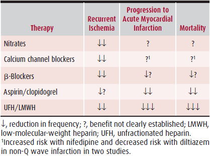
A. NITRATES—Nitrates are generally considered one of the cornerstones of therapy (Table 7–3); however, their use is largely based on clinical experience, not on randomized clinical trials. Nitrates relieve and prevent ischemia by improving subendocardial blood flow in the ischemic zone through their vasodilator actions, predominantly on the large epicardial vessels, including the stenotic segments and the coronary collaterals. Nitrates directly stimulate cyclic guano-sine monophosphate (GMP) production, achieving vasodila-tory effect in the vascular smooth muscle without requiring an intact or functional endothelium; hence, their effects are generally well preserved in atherosclerosis. Reduction of left ventricular preload and afterload by peripheral vasodilator actions may contribute to the reduction of myocardial ischemia. Although nitrates may reduce the number of both symptomatic and asymptomatic episodes of myocardial ischemia in unstable angina, no effect has yet been demonstrated on the incidence of progression to MI or death.
Table 7–3. Effects of Medical Therapy in USA/NSTEMI

Stay updated, free articles. Join our Telegram channel

Full access? Get Clinical Tree


