Fig. 42.1
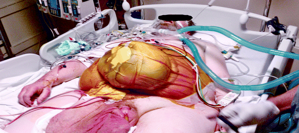
Global- and close-up view of modern intensive care unit suite. Note space limitations at the head of the bed in the first image

Fig. 42.2
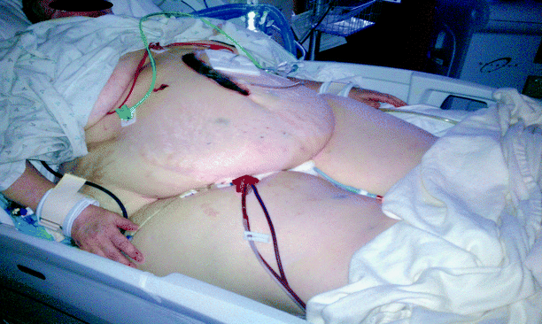
Same obese male s/p incarcerated inguinal hernia repair requiring paralysis for open abdominal decompression postoperatively. Pt is high risk for DVT due to immobility and anticoagulation is contraindicated given his recent operation

Fig. 42.3
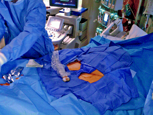
A morbidly obese patient at high risk for DVT prior to bedside filter placement

Fig. 42.4
Same patient as in Fig. 42.3 after sterile preparation of the groin. Note that an assistant may be needed to retract excess skin and fat in morbidly obese patients
Transabdominal duplex ultrasound (DUS) and intravascular ultrasound (IVUS) have emerged as familiar and adaptable techniques for vena cava filter insertion (Table 42.1). Accordingly, our rapid advances in diagnostic imaging quality have allowed us to push the envelope of making filter placement less invasive to the patient. It is natural to think that this technology will eventually be so robust that it makes bedside placement the new norm in IVC filter placement.
Table 42.1
Series of ultrasound-guided vena cava filter placement
Author | Year | N | Modality | Location | Technical success (in %) | Misplacement (in %) | Overall complication rate (in %) |
|---|---|---|---|---|---|---|---|
Killingsworth [2] | 2010 | 109 | IVUS | Bedside | 97 | 6 | 5 |
Aidinian [4] | 2009 | 14 | IVUS | Bedside | 100 | 0 | 0 |
Spaniolas [3] | 2008 | 47 | IVUS | IR/Angio/OR | 100 | 2 | 15 |
Kardys [5] | 2008 | 31 | IVUS | OR | 97 | 3 | 3 |
Corriere [9] | 2005 | 382 | DUS | Bedside | 97 | 5 | 2 |
Rosenthal [10] | 2004 | 94 | IVUS | Bedside | 97 | 3 | 6 |
Garrett [11] | 2004 | 28 | IVUS | Bedside | 93 | 8 | 15 |
Gamblin [12] | 2003 | 36 | IVUS | OR | 94 | 0 | 0 |
Wellons [13] | 2003 | 45 | IVUS | IR/bedside | 94 | 3 | 6 |
Conners [14] | 2002 | 284 | DUS | Bedside | 98 | 2 | 4 |
Ashley [15] | 2001 | 21 | IVUS | OR | 100 | 0 | 0 |
Ebaugh [16] | 2001 | 26 | IVUS | Bedside | 92 | 4 | 12 |
Bonn [17] | 1999 | 30 | IVUS | IR Suite | 100 | 0 | 0 |
Sato [18] | 1999 | 53 | DUS | Bedside | 98 | 0 | 2 |
Pulmonary embolism reduces the cross-sectional area of the pulmonary vascular bed, resulting in an incremental increase in pulmonary vascular resistance and subsequent increased right ventricular afterload [24]. In its most severe form, cardiopulmonary collapse ensues leading to death. Ten percent of patients who develop pulmonary embolism die within the first hour, and 30% die subsequently from recurrent embolism, making prevention and prompt recognition, when prevention fails, a priority [25]. Anticoagulation alone can decrease the mortality rate to less than 5%, but cannot be used universally in all patient populations. Thus, absolute and relative indications for IVC filter placement include, and are largely unchanged (Table 42.2).
Table 42.2
Indications for IVC filter placement
Absolute indications | Documented VTE w/contraindications to anticoagulation* |
Documented VTE despite adequate anticoagulation | |
Complications arising from anticoagulation | |
Concurrent pulmonary embolectomy | |
Failure of alternate caval interruption methods | |
Relative indications | Proximal free-floating iliofemoral thrombus (>5-cm-free-floating tail) |
Propagation of an iliofemoral thrombus despite therapeutic anticoagulation | |
VTE in certain high-risk populations including those with severely compromised cardiopulmonary function (e.g., severe pulmonary hypertension) undergoing high-risk operations (e.g., spine or bariatric operations) |
Absolute Indications
Generally accepted absolute indications for IVC filter insertion include:
1.
Contraindications to anticoagulation*
2.
Documented VTE while in the therapeutic anticoagulation range
3.
Complications arising from anticoagulation
4.
Concurrent pulmonary embolectomy
5.
Failure of alternate caval interruption methods
*Examples of contraindications to anticoagulation include clinical evidence of active or recent hemorrhage (i.e., intracranial, gastrointestinal) recent or planned major operation, or severe traumatic injuries.
Relative Indications
Traditional relative indications for IVC filter placement include:
1.
Detection of a proximal free-floating iliofemoral thrombus (>5 cm free-floating tail)
2.
Propagation of an iliofemoral thrombus despite therapeutic anticoagulation
Prophylactic Indications
Prophylaxis against VTE remains the most common indication for IVC filter insertion in most modern series [22]. While not supported by randomized trials, a growing clinical experience drives prophylactic filter placement following trauma in patients at highest risk for PE including severe head injury and coma, spinal cord injuries with neurological deficit, spine fractures with immobility, pelvic and multiple long bone fractures, and perhaps, direct venous trauma. Other recommended prophylactic indications include malignancy, hemorrhagic stroke, recent neurological surgery, or concurrent open bariatric surgery [15–18].
Technique
Equipment
The bedside filter insertion techniques that are described below have been developed through extensive clinical experience and have been optimized with the following equipment: Stainless Steel Over-the-Wire Greenfield Vena Cava Filter (Boston Scientific, Natick, MA), Gunther Tulip Vena Cava Filter (Cook Medical, Bloomington, IN), a portable duplex ultrasound system (Philips Medical Systems, Andover, MA), and a portable IVUS imaging system (Galaxy IVUS imaging system with an Atlantis PV Peripheral Imaging Catheter, 15 MHz, SF profile, Boston Scientific, Natick, MA). Although our experience with ultrasound-guided cava filter insertion has been dominantly accomplished with the retrievable Gunther Tulip filter and to a lesser extent the stainless steel Greenfield filter, the described techniques can be readily applied to placement of other commercially available IVC filters.
Technique for Transabdominal Duplex Ultrasound
Preprocedural Imaging
Our preferred technique for both transabdominal duplex and intravascular ultrasound guided filter placement has been previously described [23]. Though not always available, any CT images of the abdomen are reviewed to allow preprocedural determination of the IVC filter diameter. Initial transabdominal duplex ultrasound is performed to define the suitability of bedside filter placement under ultrasound guidance. The inferior vena cava is interrogated transversely and longitudinally at the renal vein confluence. Adequate visualization of a renal vein (or right renal artery) is mandatory to guide accurate filter insertion. Before proceeding, it is important to confirm an IVC diameter <28 mm, and patency of the iliocaval and femoral venous systems, respectively. Visualization of intraluminal thrombus should steer the clinician toward conversion to conventional cavography under fluoroscopic guidance. Alternate central venous access, such as the internal jugular or brachial, in the setting of bilateral common femoral or iliac venous thromboses is possible, but routine use of the internal jugular vein is discouraged due to the space limitations inherent to critical care rooms and achieving wire access to the inferior vena cava without fluoroscopic guidance can be challenging (Fig. 42.1).
Filter Placement Technique
After the administration of a local anesthetic, ultrasound-guided seldinger technique is used to cannulate the common femoral vein and place a 0.035-in. guide-wire into the IVC. The device delivery sheath is inserted to the level of the renal vein-IVC confluence. The delivery catheter containing the preloaded IVC filter (whether permanent or temporary) is placed into the sheath and advanced to the tip of the sheath. When implanting a Greenfield filter, the wire should be removed to precisely identify the tip of the filter. Once the renal vein-IVC confluence is clearly recognized in the transverse orientation, the sheath and filter are positioned caudal to the renal vein ostium. At this stage, the ideal deployment position is reached. When visualized longitudinally, the filter should be easily recognized and its tip should approach the right renal artery, a reliable landmark for the renal vein-IVC confluence. The filter is then deployed under continuous ultrasound guided visualization.
Completion Imaging
After deployment, correct filter position, complete leg expansion, and presence/lack of tilt are confirmed via immediate transabdominal ultrasound and subsequent abdominal plain film. Gentle manual compression to the site where the device entered the vein (as opposed to the needle mark in the skin) provides rapid and lasting hemostasis.
Technique for Intravascular Ultrasound
Preprocedural Imaging
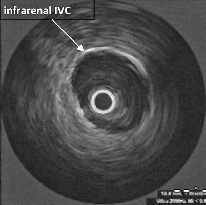
The location and patency of the common femoral veins at the intended venous puncture sites are confirmed with duplex ultrasonography, similar to the transabdominal approach. Then, the puncture site and surrounding 1 cm of the tissue is infiltrated with local anesthesia. The common femoral vein is cannulated and a guidewire is inserted into the IVC percutaneously. An 8-Fr sheath is then inserted into the common femoral vein through which the IVUS probe is advanced into the IVC to the level of the right atrium. Venous anatomy is sequentially identified during a “pull-back” technique including the right atrium, hepatic veins, renal veins, infrarenal IVC, and iliac venous confluence (Figs. 42.5, 42.6, 42.7, 42.8, and 42.9). The IVUS catheter is withdrawn to a site immediately caudad to the lowest renal vein and the IVC diameter is determined.


