Fig. 17.1
Case tricuspid regurgitation auscultation diagram. Auscultation demonstrated a holosystolic murmur with a normal S2 heard at the lower left sternal border
The liver is non-pulsatile.
Test Results
Electrocardiogram demonstrates sinus rhythm, with a rightward axis, and RSR’ pattern suggestive of incomplete RBBB (Fig. 17.2).

Fig. 17.2
12-lead echocardiogram showing an incomplete right bundle branch block pattern with rightward axis
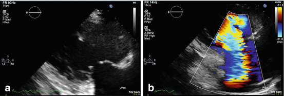
Echocardiogram (Fig. 17.3a, b) showed a severely depressed ejection fraction (EF) and right ventricular enlargement (RVE) with decreased right ventricular ejection fraction (RVEF). Due to RVE, there was incomplete coaptation of the valve leaflets and severe tricuspid regurgitation (TR) with a peak velocity of 3.4 m/s. The IVC demonstrated dilation without respiratory collapse. Right ventricular systolic pressure (RVSP) is estimated to be 60–65 mmHg.

Fig. 17.3
(a, b) Case echocardiograms. (a) 2-dimensional echocardiogram of the tricuspid valve in systole showing significant leaflet separation (malcoaptation) in systole. (b) Color-Doppler echocardiogram in the same projection showing severe tricuspid regurgitation through the separated valve leaflets
Clinical Basics [1]
Normal Anatomy
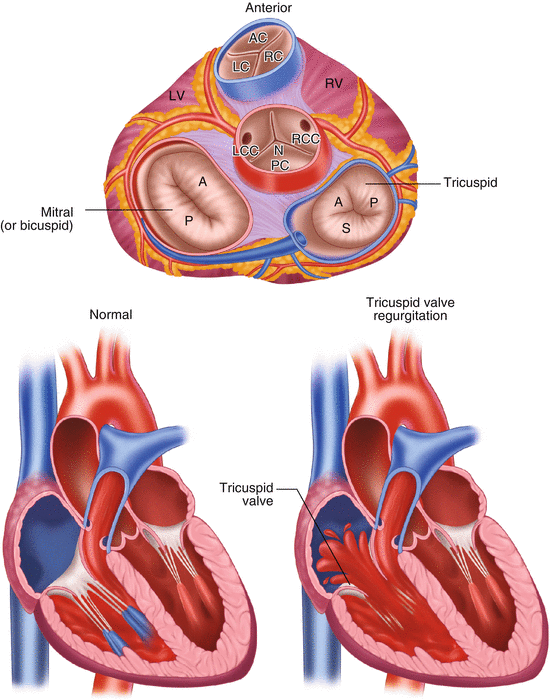
The tricuspid valve is composed of three leaflets: septal, anterosuperior, and inferior leaflets (Fig. 17.4).

Fig. 17.4
Normal anatomy of tricuspid valve. Tricuspid valve is made up of three leaflets connected to the tricuspid annulus. In tricuspid regurgitation blood will flow from the right ventricle into the right atrium due to insufficiency of the valve. The insufficiency can be due to a direct insult to the valve itself (congenital abnormalities, infective endocarditis) or secondary to right ventricular dilation and annulus dilation
Definitions
Tricuspid regurgitation is defined by retrograde blood flow from the right ventricle to the right atrium due to an insufficient valve.
Etiology (Table 17.1)
Table 17.1
Primary and secondary causes of tricuspid regurgitation
Primary causes of TR | Secondary causes of TR |
|---|---|
Congenital valve abnormalities – Ebstein anomaly, Malpositioned TV annulus | Pulmonary hypertension |
Rheumatic heart disease | Right ventricular failure |
Carcinoid heart disease | Right ventricular infarction |
Endomyocardial fibrosis | Congenital pulmonary hypertension |
Traumatic valve injury | Left sided heart failure |
Infective endocarditis | |
Dilated cardiomyopathy | |
Papillary muscle dysfunction or valve prolapse | |
Myxomatous leaflets | |
Malpositioned TV annulus |
It can occur from primary valvular pathologies or secondary to increased RVSP, generally greater than 55 mmHg.
The most common cause of TR is left sided heart failure, which results in increased right ventricular volumes and subsequent functional TR.
Causes of functional, or secondary, TR also include:
pulmonary hypertension (congenital or acquired).
right ventricular failure or infarction.
The annulus of the valve, located in the right atrium, is commonly dilated in response to large pressures or volumes entering the heart and can be another secondary cause of regurgitation.
Intrinsic valvular disease leading to primary TR is much less common but can result from a variety of pathologies. Primary causes include:
congenital valve abnormalities.
disorders or trauma leading to papillary muscle dysfunction.
valve prolapse.
ruptured valve.
Signs and Symptoms
The signs and symptoms of TR vary depending on the severity of the insufficiency but can include ascites, cyanosis, jaundice, hepatomegaly, edema, fatigue, and dyspnea.
Other clinical findings may be a systolic wave in the venous pulse, jugular venous distention with large v waves, an impulse in the right ventricle, systolic pulsations in the eyeballs and pulsations in the neck.
A systolic murmur is often heard on auscultation and is an important clinical finding and useful diagnostic and prognostic marker.
TR is generally heard best at the left lower sternal border but may also be loudest in the epigastrium or right upper sternal border.< div class='tao-gold-member'>Only gold members can continue reading. Log In or Register to continue

Stay updated, free articles. Join our Telegram channel

Full access? Get Clinical Tree


