Transthoracic Resection of the Esophagus
Mark S. Allen
Esophageal resection is most frequently performed for carcinoma of the esophagus. However, esophagectomy is also occasionally necessary in the treatment of some benign esophageal diseases, such as irreparable or neglected perforations, end-stage achalasia, intractable peptic structures, and recurrent failures after antireflux surgery.
Although esophageal carcinoma is uncommon in the United States, it is a highly lethal malignancy. The incidence of esophageal carcinoma, particularly that of adenocarcinoma, has risen dramatically during the past decade.4,5 About 15,560 people are diagnosed with esophageal cancer in the United States each year and, unfortunately, almost 14,000 of these die of the disease.14 Surgical resection remains the standard of treatment for early-stage disease, but since the survival rates are so low, multiple randomized trials have attempted to prove a survival benefit with either induction chemoradiotherapy or postoperative chemoradiation. Proving a survival benefit from these additional therapies has been difficult; thus, there is ongoing controversy regarding the preferred management.
In addition there remains controversy in the surgical community related to the best surgical approach (transhiatal or trans- thoracic) and the extent of lymphadenectomy (Table 136-1). Some surgeons regard esophageal cancer as a systemic disease at the time of diagnosis, with palliation being the essential surgical objective. In that paradigm, surgical cure is essentially a chance phenomenon. Others believe that with complete surgical resection cure is possible and advocate a complete resection of the esophagus with the draining lymphatics. This en bloc esophagectomy involves resection of the esophagus within wide envelope of adjoining mediastinal tissue accompanied by a thorough dissection of the mediastinal and upper abdominal retroperitoneal lymph nodes. More recently, further extension of the operations included dissection of lymph nodes in the superior mediastinum and lower neck. Although the 5-year survival after these extended resections has been encouraging, they remain controversial in most of western Europe and the United States, since proving there is a difference in the various surgical approaches has been difficult.22,27
Historical Background
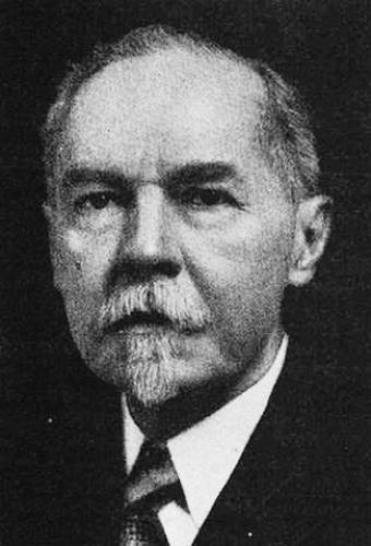
The surgical treatment of esophageal carcinoma is a relatively recent undertaking and thus has evolved rapidly over a short time span. Although partial esophageal resections were attempted in the nineteenth century, Naef23 described that early in the twentieth century, respected surgeons such as Sauerbruch declared that resection of the midesophagus was impossible. However, Franz Torek accomplished this feat on March 14, 1913 (Fig. 136-1).33 He performed an esophagectomy through the left chest with considerable efforts to dissect the esophagus behind the aortic arch and with a resultant injury to the left main bronchus, which was repaired. The patient survived with a cervical esophagostomy and gastrostomy for 13 years.
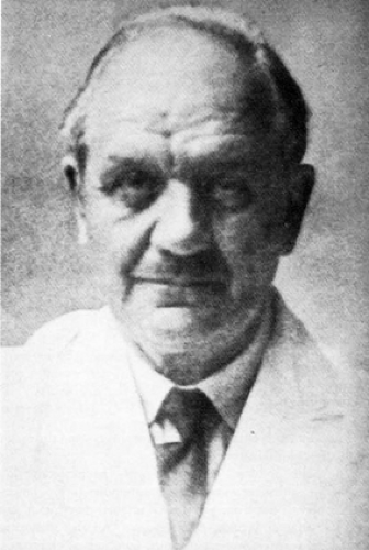
Intrathoracic restoration of esophagogastric continuity would have to wait until 1933, when the Japanese surgeon Oshawa25 and, soon after, the American surgeons Marshall19 and Adams and Phemister2 accomplished an esophagogastric anastomosis for distal esophageal carcinomas. In 1942, Churchill and Sweet8 delivered their paper entitled “Transthoracic Resection of Tumors of the Stomach and Esophagus,” which ushered in the modern era of transthoracic approaches to esophageal lesions. Garlock and Sweet successfully proceeded more proximally and performed a supra-aortic esophagogastrostomy with placement of the conduit in front of the aorta, as described by Garlock.11 Sweet became the leading authority in esophageal surgery (Fig. 136-2). His preliminary report, “Surgical Management of the Midthoracic Esophagus,” outlined techniques that have become the foundation of modern esophageal surgical teachings and are still relevant.31
The left-sided transthoracic approach to the esophagus had its shortcomings. Numerous patients had postoperative complications related to respiratory distress, which was a result of the radial incision in the diaphragm. Also, the blind dissection behind the aortic arch could be challenging. The British surgeon Ivor Lewis in 1946 proposed a right-sided approach, which allowed for dissection of the midesophagus under direct vision and did not require a diaphragmatic incision (Fig. 136-3). It did
not take long to convert thoracic surgeons to the Ivor Lewis approach for proximal and midesophageal lesions, whereas the left-sided “Sweet approach” was reserved for distal carcinomas. The fear of pleural leakage from an intrathoracic anastomosis motivated surgeons to place the esophagogastric anastomosis into the neck. McKeown21 described a technique of laparotomy, right thoracotomy, and cervical anastomosis.
not take long to convert thoracic surgeons to the Ivor Lewis approach for proximal and midesophageal lesions, whereas the left-sided “Sweet approach” was reserved for distal carcinomas. The fear of pleural leakage from an intrathoracic anastomosis motivated surgeons to place the esophagogastric anastomosis into the neck. McKeown21 described a technique of laparotomy, right thoracotomy, and cervical anastomosis.
Table 136-1 Types of Esophageal Resection | |
|---|---|
|
 Figure 136-1. Dr. Frank Torek. Credited with the first transthoracic esophageal resection, performed in 1913. |
 Figure 136-2. Dr. Richard Sweet. Developed the basis for the modern approach to transthoracic esophagectomies with Dr. Churchill at the Massachusetts General Hospital in the 1940s and 1950s. |
 Figure 136-3. Ivor Lewis. A British surgeon who performed the two-stage esophagectomy via an abdominal approach and subsequent right thoracotomy that bears his name today. |
Table 136-2 Mortality, Morbidity, and Survival in Recent Series | ||||||||||||||||||||||||||||||||||||||||||||||||||||||||||||||||||||||||||||||||||||||||||||||||||||||||||||||||||
|---|---|---|---|---|---|---|---|---|---|---|---|---|---|---|---|---|---|---|---|---|---|---|---|---|---|---|---|---|---|---|---|---|---|---|---|---|---|---|---|---|---|---|---|---|---|---|---|---|---|---|---|---|---|---|---|---|---|---|---|---|---|---|---|---|---|---|---|---|---|---|---|---|---|---|---|---|---|---|---|---|---|---|---|---|---|---|---|---|---|---|---|---|---|---|---|---|---|---|---|---|---|---|---|---|---|---|---|---|---|---|---|---|---|---|
| ||||||||||||||||||||||||||||||||||||||||||||||||||||||||||||||||||||||||||||||||||||||||||||||||||||||||||||||||||
Stay updated, free articles. Join our Telegram channel

Full access? Get Clinical Tree


