INDICATIONS/CONTRAINDICATIONS
Because many patients with Zenker’s diverticula are elderly, an endoscopic approach offers potential advantages by avoiding neck incision. However, several technical points need to be emphasized. It is important to divide the cricopharyngeus muscle completely, and the diverticulum should be at least 3 cm in size if considering the transoral approach to allow for entrance of the stapler and a complete cricopharyngeal myotomy. Significantly larger diverticula (>6 cm) are also a relative contraindication when considering a transoral approach, particularly when the diverticulum deviates markedly to one side, or when there are bilateral pouches, as this approach may leave a considerable residual diverticulum. Other relative contraindications include the presence of prominent upper incisors, limited mouth opening, and the inability to adequately extend the neck; these anatomic features make an endoscopic repair technically difficult.
 PREOPERATIVE PLANNING
All patients should be evaluated by a barium swallow and upper gastrointestinal esophagoscopy. The Zenker’s diverticulum should be at least 3 cm in size to allow a complete transoral cricopharyngeal myotomy. In addition, patients should be evaluated for limited mouth opening and inability to adequately extend the neck, which can make placement of the Weerda laryngoscope difficult. An esophagogastroduodenoscopy (EGD) should be performed at the time of the procedure by the operating surgeon (even if it has been done preoperatively) to evaluate the diverticulum, remove any debris from the diverticulum, and examine the esophagus for any other pathology. During flexible endoscopy, the anatomy and entry into the native esophagus can be challenging. If there is difficulty in identifying the normal anatomy, we place a flexible guidewire into the native esophageal lumen and advance this into the stomach. This facilitates identifying the anatomy when inserting of the Weerdascope.
 SURGERY
Necessary equipment for a transoral Zenker’s diverticulectomy includes the following.
 Weerda laryngoscope (Karl Storz, Tuttlingen, Germany)
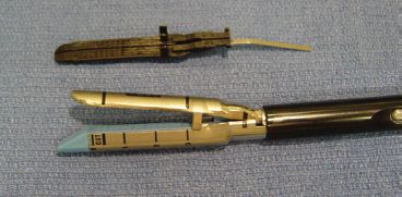
 Low-profile endostapler to allow stapling and cutting of the sphincter muscle to the end of the stapler (Fig. 19.1)
 5-mm 30-degree laparoscope to allow for additional visualization
 0.035 flexible guidewire
Positioning
Patient is placed supine on the operating table with the head positioned at the top of the operating table. General anesthesia is established using a small diameter (7 mm) endotracheal tube. Arms are typically tucked at the patient’s sides. The operating table should be rotated so that the anesthesia team is to the left.
Technique
 With the patient adequately anesthetized, flexible esophagoscopy is performed to assess the diverticulum and also to suction any retained material from the diverticulum. At this time, we sometimes place a guidewire in to the native esophagus to help us place the Weerdascope properly.

Figure 19.1 Stapler with anvil.

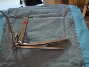
Figure 19.2 Weerda laryngoscope. (With kind permission from: Morse CR, Fernando HC, Ferson PF, et al. Figure 1: Preliminary experience by a thoracic service with endoscopic transoral stapling of cervical (Zenker’s) diverticulum. J Gastrointest Surg. 2007;11:1901–1904, Springer Science+Business Media).
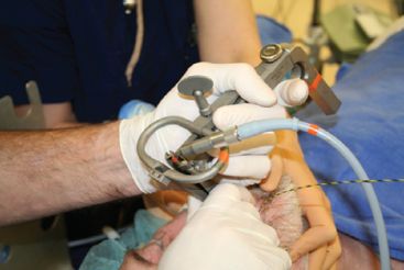
 Rigid esophagoscopy is performed using the Weerda laryngoscope (Karl Storz, Tuttlingen, Germany). This scope has two jaws (Figs. 19.2 and 19.3). The upper jaw, which is longer, is placed into the native esophageal lumen and the second is placed in the diverticulum. The jaws are then expanded which allows clear visualization of the diverticulum and the common septum formed by the cricopharyngeus muscle and the esophagus.
 The placement of a 5-mm 30-degree laparoscope alongside the Weerda laryngoscope can further enable visualization of the distal anatomy during stapler placement and firing.
 A traction suture (US Surgical Endostitch, Norwalk, Connecticut) is placed in the common septum (Fig. 19.4). The traction suture helps to expose the septum and facilitates a complete cricopharyngeal myotomy.
 Using the suture to provide proximal traction on the common septum, the stapler is placed across the septum and fired (Fig. 19.5). When positioning the stapler, the flat part of the stapler is placed within the diverticulum, and the disposable cartridge is placed within the esophageal lumen. Further firings of the stapler are performed as needed to ensure the common septum is divided to the base of the diverticulum. Occasionally, the traction stitch is replaced after a firing of the stapler to further expose any residual septum.
 It is essential to realistically evaluate the progress of the procedure, and to abandon the transoral approach if it becomes unusually difficult. It is far safer for the patient to have a well-performed open operation than a complication of an endoscopic procedure.

Figure 19.3 Placement of the Weerda laryngoscope.


