Thoracic Approaches to Esophagectomy


Thoracic Approaches to Esophagectomy


Shanda H. Blackmon

Wayne Hofstetter

Stephen G. Swisher


All images (unless otherwise noted) remain the copyright of Shanda Blackmon, MD, MPH through The Methodist Hospital, Houston, TX.


INTRODUCTION

Thoracic approaches to esophagectomy may be helpful for treating benign and malignant esophageal disease. Benign indications are less common but include undilatable, recurrent strictures; megaesophagus from achalasia, trauma, or ingestion of acids or alkali; and recurrent foregut surgery for benign processes (Table 18.1 and Fig. 18.1). More common indications include primary or salvage esophagectomy for localized esophageal cancer. Less common malignancies, such as sarcomatoid carcinoma, gastrointestinal stromal tumor, small-cell cancer, and locoregional involvement of other malignancies may also be addressed by transthoracic esophagectomy. Various thoracic approaches have been successfully used to address esophageal disorders (Table 18.2), the choice of which depends not only on surgeon preference but also on tumor location, body habitus, history of prior operations, patient condition, choice of esophageal substitute, and prior radiation therapy. A skilled esophageal surgeon does not limit the approach to a single modality but instead adopts the best approach to the individual patient. The primary goal of esophageal carcinoma treatment remains neoplasm resection, with relief of dysphagia. When selecting an approach, prior operations and conduit irradiation should be considered. In some situations such as in salvage esophagectomy, the use of a heavily irradiated conduit is unavoidable. In these cases, buttressing the esophageal anastomosis with a pedicled omental or muscle flap may be beneficial. Other challenges include previous extensive lung resections on the operated or contralateral side, which may require a left-sided thoracoabdominal approach instead of a traditional Ivor-Lewis right thoracic approach. High-risk esophageal resections can also be performed in a delayed or two-stage operation if there is concern regarding the performance of an anastomosis at the time of resection.


RESECTION

When esophageal resection is performed for malignant disease, the entire diseased esophagus, including any dysplasia, metaplasia, or cancer, should be removed. Recurrent cancer after a low intrathoracic anastomosis can be due to incomplete resection or recurrent disease. Adenocarcinoma can occur at multiple levels, and the seventh edition of the American Joint Committee on Cancer (AJCC) staging manual (Fig. 18.2) includes proximal gastric cancer in the esophageal cancer staging system. The management of type I and II gastroesophageal junction cancers includes extending the resection to the thoracic esophagus in order to obtain an optimum the proximal margin. A gastrectomy for type III gastroesophageal junction cancers should be performed by an expert thoracic surgeon because of the potential need for simultaneous esophageal resection. Patients who are treated with definitive chemoradiation therapy and develop recurrent locoregional disease should be evaluated for a salvage esophagectomy if there is no evidence of distant disease. A transthoracic approach is favored in this situation because it allows complete resection of adjacent lymph node regions and direct placement of omental or muscle flaps to buttress these high-risk anastomoses. Palliative resection for dysphagia or bypass for tracheoesophageal fistula is seldom performed because of the availability of expandable covered stents. Patients who are at high risk for surgery can often be treated in a two-stage procedure or bypass to minimize the risks of a prolonged one-stage procedure.


PRINCIPLES OF TRANSTHORACIC ESOPHAGEAL RESECTIONS AND RECONSTRUCTION


Surgical Technique: Task List

Before the patient is brought into the operating room, the entire team should understand the operative plan. An equipment checklist will decrease intraoperative delays. All members of the team should be given a list of the planned surgical steps. The task list described below was developed to define each step and provide evidence for each decision that is made along the way.


Initial Operating Room Assessment

The patient is brought into the operating room, and after the induction of general anesthesia all appropriate lines and monitors are placed. Empiric antibiotics should be administered, along with a beta blocker, when indicated. Sequential compression devices should be applied to the lower extremities, with or without pharmacologic prophylaxis. A copy of the endoscopy reports and endoscopic ultrasound, computed tomography, and positron emission tomography images should be available in the room prior to beginning the procedure.


Table 18.1 Indications for Esophagectomy


Benign


Undilatable benign stricture


Megaesophagus from achalasia


Reoperative foregut surgery after fundoplication


Acid or alkali ingestion


Trauma


Benign tumors


Unrepairable leak or fistula


Gastric conduit necrosis


Malignant


Esophageal cancer


Primary esophagectomy


Salvage esophagectomy


Sarcomatoid cancer


Locoregional involvement of miscellaneous malignancies


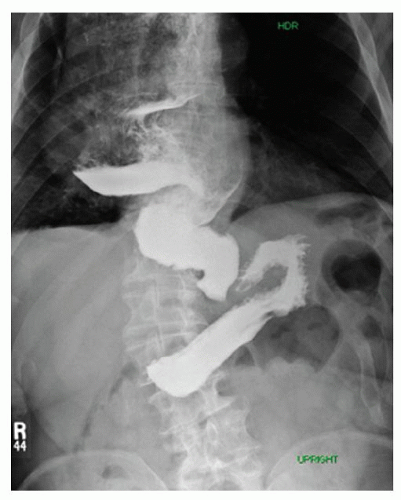
Fig. 18.1. Contrast esophagram of a sigmoid esophagus from life long achalasia.


Bronchoscopy

A bronchoscopy should be performed in all patients with tumors that occur in close proximity with the airway. The bronchoscope is advanced down to the level of the subsegmental bronchi, and the entire tracheobronchial tree is visualized for evidence of abnormalities. The absence of tracheal or left main bronchus involvement should be documented before proceeding with esophagectomy. The single-lumen endotracheal tube or laryngeal mask airway (LMA) is then removed, and a double-lumen endotracheal tube is positioned. A repeat bronchoscopy confirms that the left-sided endotracheal tube is in the left mainstem bronchus, and the tracheal opening on the right side allows for visualization of the carina and the entire right mainstem bronchus. A systems check should include monitoring the pressure in the bronchial cuff to ensure that it is not too high.


Table 18.2 Transthoracic Resections


Ivor-Lewis esophagectomy = right thoracotomy + laparotomy


Hybrid esophagectomy


  1. Laparoscopic abdominal approach + thoracotomy


  2. Laparotomy + thoracoscopic approach


  3. a or b + neck anastomosis


Minimally invasive esophagectomy


  1. Laparoscopic abdominal approach + thoracoscopic approach


  2. a + neck anastomosis


McKeown esophagectomy = three-field esophagectomy


Abdominal incision + thoracic incision + neck incision


Left-sided thoracoabdominal esophagectomy


Isolated segmental thoracic esophagectomy (rarely performed)


Endoscopy

Esophagoscopy should be performed to identify the proximal aspect of the tumor and any abnormal tissue near the tumor, including Barrett’s metaplasia. As long as the tumor is distal to 30 cm from the incisors, an Ivor-Lewis esophagectomy is possible, with an intrathoracic anastomosis. No abnormalities including metaplasia should be visible proximal to the area of planned transaction. Once the endoscopy confirms normal squamous lining proximal to 30 cm from the incisors, the patient can be positioned supine, with the abdomen sterilely prepared and draped.


Abdominal Approach

A laparoscopic abdominal approach or open midline incisional approach can be used. The advantage of an initial laparoscopic approach is detection of potentially subclinical metastatic peritoneal or liver disease prior to performing a more invasive incision (Fig. 18.3).


Laparoscopic Approach

In the laparoscopic approach, five laparoscopic abdominal trocars are placed: a 12-mm trocar in the midline or just to the right of midline and two 5-mm trocars on either subcostal area (Fig. 18.4). The right subcostal trocar is used to place a Mediflex liver retractor to retract the liver anteriorly and laterally to the right. An energy source is typically placed through the 12-mm port. A 5- to 10-mm trocar should be placed for the camera in the medial subcostal area, approximately 4 to 5 cm inferior to the subcostal region. After all trocars have been placed safely, abdominal exploration should be performed. Checking for evidence of bowel injury or metastatic disease is necessary before proceeding with the esophagectomy. A minimally invasive approach should only be used if the exact same resection quality can be achieved as in an open approach.


Open Midline Incision

An open abdominal incisional approach can typically be performed through an upper midline laparotomy incision. Alternatively, but less commonly, a chevron or thoracoabdominal incision may be employed. The midline incision extends from the xiphoid down to just above the umbilicus. When open, a self-retaining type of retractor may be used to retract the liver and adjacent costal margin out of the way. A left thoracoabdominal incision is advantageous when the patient has undergone extensive foregut surgery and has excessive scar tissue in the left upper quadrant.


Mobilizing the Esophagus

An energy source is used to divide the pars flaccida or hepatogastric ligament. The left triangular ligament should be divided to allow the left lateral segment of the liver to be retracted. The right crus of the diaphragm should be exposed with an energy device, and the esophagus should be mobilized away from the right crus, stripping the peritoneal lining away to include all tissues. If the patient has a small hiatus, the crus can be divided approximately 1 to 2 cm to enlarge the hiatus for additional dissection. This allows for circumferential dissection around
the esophagus. The phrenoesophageal membrane should be taken down, and all tissue adjacent to the esophagus should be kept with the esophagus. The crural decussation should be dissected posteriorly, and the left crus should be identified from the right side of the abdomen (Fig. 18.5). The posterior esophageal window should be developed, and all tissues adjacent to the esophagus should be dissected away from the right and left crura. After dissecting in the cranial direction toward the mediastinum, the gastrohepatic ligament should be mobilized toward the lesser curve of the stomach. An appropriately sized hiatus for conduit passage is important as too large a hiatus could result in colon herniation and too tight a hiatus can result in ischemia of the conduit. It is helpful to dissect as far into the chest as is safe before closing the abdomen, as the inferior pulmonary ligament lymph nodes from both sides can be dissected from the abdominal approach. It is usually possible to reach up to the inferior pulmonary vein, depending on patient anatomy.


Fig. 18.2. Seventh edition of the AJCC cancer staging system. (From Edge SB, Byrd DR, Compton CC, Fritz AG, Greene FL, Trotti A, eds. AJCC Cancer Staging Manual, 7th ed. Springer; 2009.)


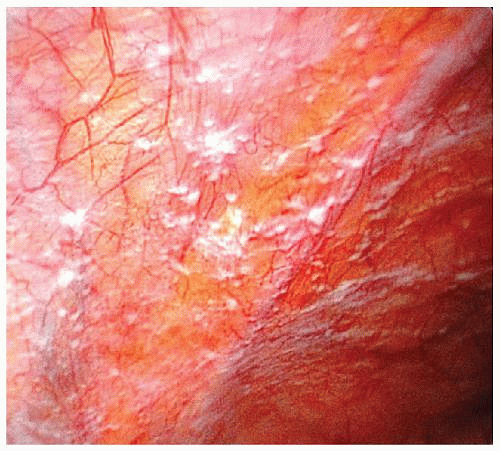
Fig. 18.3. Laparoscopic view of diffuse metastatic abdominal disease.


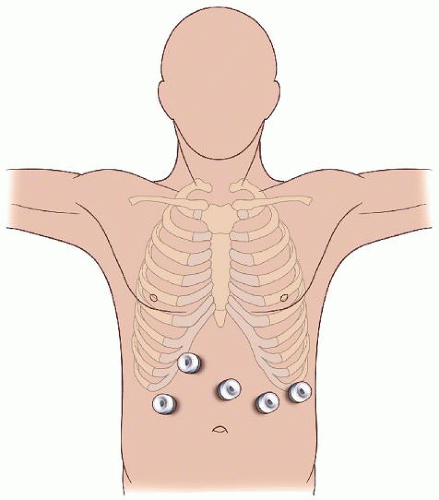
Fig. 18.4. One approach to laparoscopic trocar port placement.


Mobilizing the Stomach

The right gastroepiploic artery should be identified and preserved as the gastrocolic omentum is divided and the lesser
sac is entered (see Fig. 18.6, “gastroepiploic artery preservation and omental dissection”). Instead of sparing a small segment of omentum, a larger omental flap can be created to be delivered into the chest to cover the anastomosis (Fig. 18.7). When manipulating the stomach, care should be taken to avoid injury when grasping and the right gastroepiploic artery should be avoided. The division should be completed along the entire greater curve of the stomach up to where the short gastric vessels are encountered. Exposure of the short gastric vessels is enhanced by grasping the posterior aspect of the stomach and rolling the tissue toward the liver and up away from the aorta. The short gastric vessels should be divided with an energy device up to the left crus of the diaphragm. The posterior attachments of the stomach to the pancreas should be divided. The cardia of the stomach should be dissected away from the left crus. The posterior esophageal window should be created, and a Penrose drain placed around the esophagogastric junction. This drain can later be recaptured and pulled into the chest to assist in mobilizing the intrathoracic esophagus. The posterior aspect of the stomach should be dissected away from the pancreas, preserving the right gastric artery. For benign disease, the modification does not typically require a complete esophagectomy or lymphadenectomy.


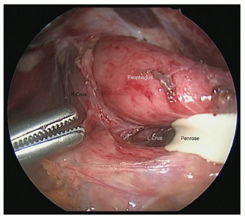
Fig. 18.5. Development of the posterior esophageal window.


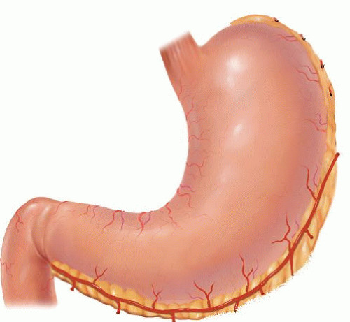
Fig. 18.6. Gastric mobilization with gastroepiploic artery preservation.


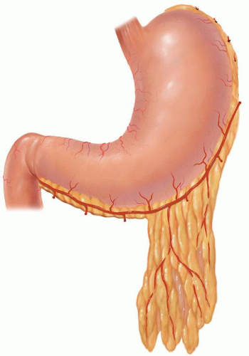
Fig. 18.7. Pedicled omental flap.


LEFT GASTRIC ARTERY DIVISION AND CELIAC LYMPH NODE DISSECTION

Once the posterior attachments of the stomach have been divided, the stomach is lifted anteriorly to allow for visualization of the left gastric artery pedicle. The left gastric artery pedicle should be divided either with individual ligation of the artery and vein or with a linear vascular stapler at the level of its origin from the celiac axis taking care to include the lymph node packet.

One should be aware for the presence of any aberrant celiac, gastric, or left hepatic artery anatomy (Fig. 18.8A, 18.8B), especially a replaced left hepatic artery coming completely from the left gastric artery. If this artery is an accessory artery and another left hepatic artery is present, it can typically be divided without consequence. However, if the entire left hepatic artery originates from a left gastric artery, a laparoscopic or open sonographic evaluation of flow dynamics can be used to determine portal flow and compensate when hepatic arterial flow has stopped. If blood flow to the liver is not diminished by more than 50%, the artery can still be divided. If the artery must be preserved, the left gastric artery should be ligated further distally, that is closer to the stomach. This will result in a lesser lymph node acquisition but will prevent hepatic necrosis. A low stapler division of this artery, as proximal as possible, allows for incorporation of all lymphatic tissue to be included with the specimen. Celiac lymph nodes can be dissected at this time and sent as separate pathologic specimens. Left gastric artery lymph nodes are then identified and dissected away from the specimen and identified for permanent specimen pathologic analysis. We recommend sending separately labeled packages to more accurately stage disease.


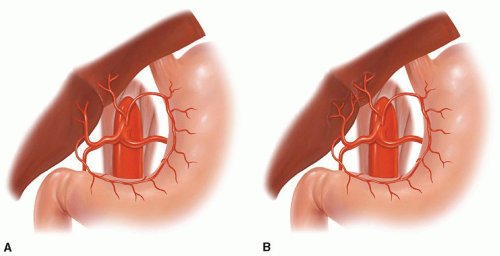
Fig. 18.8. Aberrant celiac, left gastric, or hepatic artery anatomy. (A) Replaced left hepatic artery and (B

Only gold members can continue reading. Log In or Register to continue

Stay updated, free articles. Join our Telegram channel

Jun 15, 2016 | Posted by in CARDIAC SURGERY | Comments Off on Thoracic Approaches to Esophagectomy

Full access? Get Clinical Tree

Get Clinical Tree app for offline access