Introduction to the Rhythms
 Introduction to the Rhythms
Naming Rhythms
Electrocardiograms (ECGs) are named for the pattern they represent. To name rhythms successfully, you’ll need the following vocabulary:
atrial—originating in the atria
-cardia—pertaining to the heart
compensatory—indicating a pause when the heart holds a beat
contraction—“squeezing” of the heart muscle prompted by the conduction of an electrical impulse
ectopic—occurring in an abnormal position or place
irregular—an unequal R-R interval
premature—a complex that appears earlier than expected
sinus—a normal configuration of the P wave, QRS complex, and T wave
Naming rhythms is like being a detective in a mystery novel. All the clues are right there on the strips. Here are your first three steps:
Clues to Identifying Rhythms
The best approach to identifying rhythms is to use a systematic method. One such system is presented in Cohn’s Pocket Guide to Adult ECG Interpretation. You should use this guide each time you identify a rhythm.
The following are additional pointers for recognizing ECG patterns:
Normal Sinus Rhythm
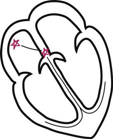
In normal sinus rhythm, the impulse follows a normal path of conduction: from the sinoatrial (SA) node to the AV node, to the bundle of His, through the left and right bundle branches, to the Purkinje fibers. The following tracing reflects a normal rate (60-100) and regular rhythm (regular R to R interval).*

Sinus Arrhythmia
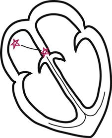
In sinus arrhythmia, a normal sinus rhythm has an irregular R-R interval. All the complexes are present in the correct order and in correct relationship to one another, but the R-R interval is varied or irregular. This rhythm is seen in some people as a result of normal breathing—nothing to get excited about.

Sinus Bradycardia
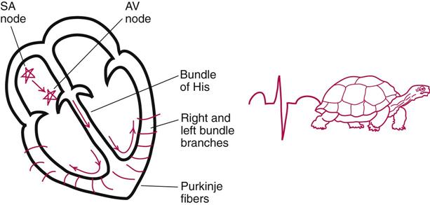
In sinus bradycardia, all the complexes are present and upright. Each complex is complete, and each interval is normal. However, the rate is below 60 beats per minute. In a young, healthy individual, this is the sign of a well-trained athlete. In an older person who is not in top physical condition, it can be the first indicator of a more serious cardiac situation.

Sinus Tachycardia
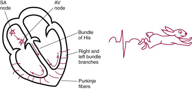
In sinus tachycardia, all the complexes are present and upright. Each complex is complete, and each interval is normal. However, the rate is above 100 beats per minute. Sinus tachycardia can be the result of fright, fever, low oxygen states (hypoxia) or pain. Sinus tachycardia also can signal a cardiac event.

Atrial Flutter
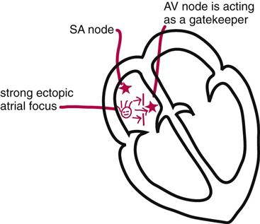
In atrial flutter, a single strong ectopic focus in the atrium starts to beat fast, 240-360 beats per minute. The AV node acts as a gatekeeper in this rhythm, allowing only some impulses through to the ventricles. Some people call it a “sawtooth pattern”; if you hold it upside down, it looks like the teeth of a saw or a serrated knife blade. No one calls it a “serrated knife blade pattern,” though—I just added that in case you grew up in the city and haven’t had any direct experience with a saw.

Atrial Fibrillation
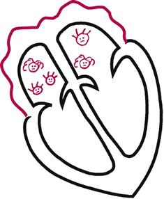
In atrial fibrillation, many weak ectopic foci in the atria beat in an uncoordinated pattern, resulting in an uneven baseline of many tiny P waves, known as fibrillatory (f) waves, in the atria. Eventually, the ventricles receive enough electrical stimulation from the atria to contract, or they contract on their own. This rhythm is characterized by a coarse baseline and an irregular distance between the QRS complexes.
Stay updated, free articles. Join our Telegram channel

Full access? Get Clinical Tree


