The ECG
 The ECG
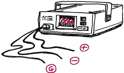
The electrocardiogram (ECG) monitor records electrical conduction through the heart. Electrical conduction is recorded through electrode pads, which adhere to the patient’s chest with a sticky gel. These electrode pads can detect the electrical activity generated by the myocardial cells. Cables attach the electrodes to an ECG monitor. A three-lead ECG monitor uses one positive electrode, one negative electrode, and one ground electrode; these often are white, black, and red, respectively (although this is not a universal system). Some ECG leads are embossed with RA for right arm, LA for left arm, and LL for left leg. Also available are 5-lead and 12-lead ECGs discussed later in the book.
Lead Placement and Vectors
The electrodes are placed on the patient so that there is one electrode on each side of the chest under the clavicles and one on the lower left side of the body.
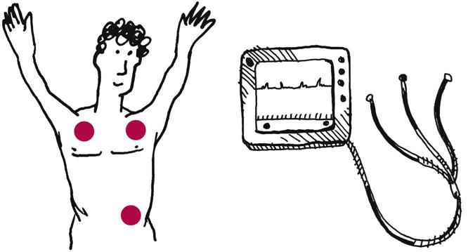
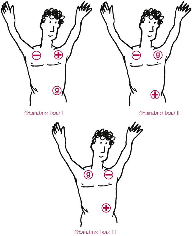
The monitor can be set to view the heart in lead I, lead II, or lead III.
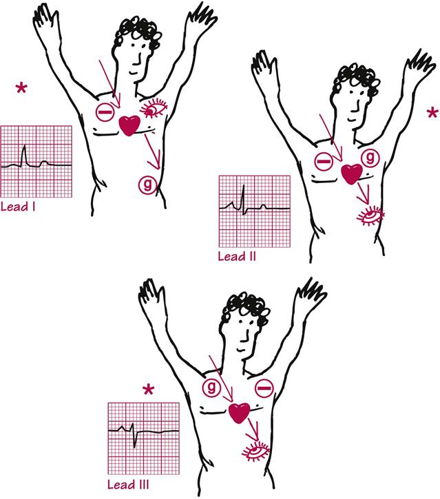
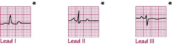
*The same complex seen in three different leads.
Settings on the machine determine which electrode is positive, which electrode is negative, and which is the ground. The positive electrode is called the “eye,” because the ECG machine is looking through the positive electrode at the electrical impulses as they travel through the heart. The ECG machine can be set to lead I, II, or lead III, and that will change which electrode is positive, negative, and ground.

Two types of electrical events affect the waveform of the ECG.
The first is the assignment of the negative and positive electrode (made by selecting lead I, II, or III on the ECG machine), because energy is recorded from the negative electrode to the positive electrode.
The second is the negative to positive depolarization of the cells of the heart. Electrical impulses start in the top right side of the heart (the right atrium), and proceed downward; as a result the heart contracts from top to bottom, and blood is pushed out of the left ventricle at the bottom (apex) of the heart (shown on p. 4).
When you choose to look at the heart in lead I, lead II, or lead III, you are changing the angle of your picture; therefore, a “normal” complex on the ECG would appear different in each of these different leads. If the heart were an exit sign, the following figure shows how it would look in lead I, lead II, and lead III. All of these views would represent a normal picture of an exit sign taken from different angles. Similarly, each lead shows a normal impulse as it travels through the heart, but it is viewed from different angles.

Electrical activity moving toward the “eye” electrode is viewed as upright complexes on the monitor. The image on the ECG monitor is like the view function of a camera. The electrical activity in the heart remains the same, but the picture changes depending on where in the heart you are taking the picture.
Two considerations are important when choosing a lead for monitoring:
1 Is there a specific part of the heart I want to watch?
2 Can I get upright complexes with a smooth, stable baseline?
Stay updated, free articles. Join our Telegram channel

Full access? Get Clinical Tree


