Transvenous pacing leads have been implicated in tricuspid valve dysfunction, and our group has adopted routine use of His bundle pacing to mitigate this effect. Three-dimensional (3D) printing technology holds great promise for advancing medicine, but the high start-up costs can be a deterrent. Seeking confirmation of optimal lead placement relative to the tricuspid annulus, we used low-cost commercial and public domain technologies to generate 3D-printed hearts from selected patients with His bundle pacing leads. Our models successfully demonstrated that such lead placements avoided interference with the tricuspid valve apparatus in these cases. Future applications of 3D printing include facilitating research to minimize lead-valve interactions, understand complex cardiac anatomy, and plan complex surgical procedures.
A recent publication noted that physical three-dimensional (3D) models in the hands of students are superior to displays in books or on computers when learning anatomic concepts. We reviewed clinically indicated cardiac computed tomography (CT) scans of our patients with His bundle pacing leads and generated 3D-printed models from 2 cases, to better examine the positions of the leads relative to the tricuspid annulus.
Case Report
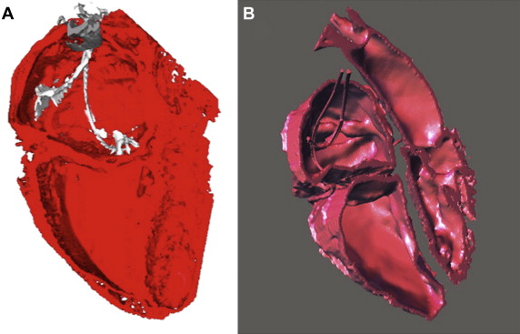
CT scans were obtained with a GE (Fairfield, Connecticut) LightSpeed 64 slice unit, with retrospective gating and a slice thickness of 0.625 mm; one was indicated for chest pain evaluation and the other before atrial fibrillation cryoablation. Models were printed with a MakerBot Replicator Mini (MakerBot, Brooklyn, New York) using polyactic acid filament and standard resolution settings. DICOM data from the CT scans were segmented with ITK-Snap ( www.itksnap.org ) as shown in Figure 1 and processed finally with MakerBot Desktop 3.0 software. Printing of each model took approximately 7 hours, and a materials cost under $10.


Stay updated, free articles. Join our Telegram channel

Full access? Get Clinical Tree


