1. Vascular lab (accredited) standards
(a) Written report that can be reviewed by an insurance company
(b) Images recorded and available for review, including anatomy, reflux, and measurements of size
(c) Cursor used for electronic measurements and recorded on the image
2. Patient examined in the standing position
3. Reflux, if in great saphenous or small saphenous, present at the junction of the superficial and deep venous system
4. Vein diameter >3 mm (some use >5 mm)
5. Reflux in vein is >0.5 s (some use >1.0 s)
Presence of Superficial Reflux
Previously, the simple presence of reflux was all that was required for authorization of an endovenous procedure, but many insurance companies now require that the length of time of the reflux be documented. Although >0.5 s is the SVS vascular lab standard for venous reflux, some health plans now require >1 s of reflux to authorize a venous procedure, based on the rationale that longer reflux time is indicative of more severe venous disease.
Location of Reflux
Although any refluxing vein can cause symptoms, most insurance authorizations require that the reflux occur at the proximal junction of the superficial vein with the deep venous system. Consequently, duplex scan reports that state that the reflux is at the saphenous-femoral junction or sapheno-popliteal junction are less likely to be denied or require a peer-to-peer review. Astute clinicians know that a refluxing distal saphenous vein can be as symptomatic as a refluxing proximal saphenous vein, but there is still a higher likelihood of insurance denial when the reflux occurs distally.
Size of the Refluxing Vein
Although any refluxing vein can cause symptoms, there are general size criteria, which vary from one insurance company to another. In the western United States, most companies require a vein to be >3 mm to justify insurance coverage and some require a vein to be >5 mm. To achieve this threshold, all patients must be evaluated in the standing position. Since veins dilate during the day and after prolonged standing, we often ask patients with marginal-sized veins to undergo their duplex ultrasound at the end of the day, and after a period of prolonged standing. Since venous symptoms occur at the end of the day, when the veins are most dilated, it seems logical to test patients when they are most symptomatic.
Role of Ultrasound During Venous Procedures
Technique
The proper use of duplex ultrasound during an endovenous ablation procedure, whether using a laser or a radiofrequency catheter, is a key to successful ablation. Duplex ultrasound competency by the vascular surgeon is critical for these procedures. An ultrasound transducer should be used to initially identify the path of the incompetent vein, for micropuncture access to the vein, during positioning of the catheter at the junction between the superficial and deep system, during infiltration of tumescence solution, and to visualize and compress the vein during the ablation procedure.
Duplex Venous Evaluations Immediately Prior to the Procedure
Some patients arrive for the ablation procedure with a duplex scan having only been performed at another facility or with incomplete information from a unaccredited vascular lab. Consequently, a duplex scan can be used by the vascular surgeon performing the ablation procedure to confirm the findings from another vascular lab and to prepare for the procedure, before opening the disposable supplies, which are expensive and would be wasted if the procedure did not need to be performed. Prior to the ablation procedure, duplex ultrasound should be used to confirm the location of the reflux, diameter of the vein(s), and then to mark the path of the vein or site of ablation. This usually requires the patient to be in a sitting or standing position, particularly if confirmation of valve reflux is needed. If the access vein for ablation procedure is small on this initial evaluation, nitroglycerine can be placed over the site of access to dilate the vein and make access easier. If a patient is fearful of needle sticks, then the entire path of the vein can be marked with duplex scan imaging and topical xylocaine placed on the skin along the path of the incompetent vein to reduce the discomfort from the needle sticks that are used for local anesthesia and tumescence.
Duplex Scan During the Procedure
Endovenous ablation procedures should be performed using a tilt table and portable or fixed duplex ultrasound.
Saphenous Ablation
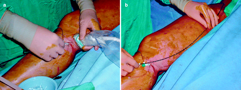
Percutaneous placement of the catheter in the below knee saphenous vein is the access site of choice (Fig. 44.1). The vein can be identified by finding the saphenous vein in the thigh, which is under the fascia in the medial thigh. If this vein is followed from the thigh to the calf, it usually is of adequate caliber for access. Once access is obtained under ultrasound guidance and the sheath has been placed, the catheter is then passed up the saphenous vein and parked 2 cm from the saphenous femoral junction and below the epigastric vein. This position is best determined using B-mode ultrasound guidance in both the transverse and saggital planes. The distance between the tip of the catheter and the junction of the superficial and deep vein should be measured with the ultrasound cursor. The relationship of the catheter to the epigastric vein is variable, so distance from the saphenofemoral junction is the most critical measurement. Liberal injection of tumescent solution, consisting of saline, xylocaine, and epinephrine, is placed around the saphenous vein from the catheter insertion site to the saphenofemoral junction, under ultrasound guidance. Although different techniques may be used to deliver tumescence, we place the transducer in a transverse position and observe the infusion ultrasonically as a spinal needle infuses the tumescent solution into the perivenous space. The vein should be moved to at least 2 cm below the skin to avoid skin burn, and the tumescence should surround the entire vein to eliminate pain when the vein is being ablated. When the patient is placed in the Trendelenberg position to minimize vein diameter during ablation, the catheter position should be rechecked by duplex scan with respect to the junction. During ablation, the vein can be visualized under ultrasound guidance, to assure that compression over the vein is directly transmitted to it.


