The Middle Mediastinum

CHAPTER 42 The Middle Mediastinum


The mediastinum is bounded laterally by the two pleural spaces. Within this space are located the tracheobronchial tree, the heart and great vessels, a portion of the gastrointestinal tract, and a portion of the lymphatic system. Superiorly the mediastinum is bordered by the thoracic inlet, and inferiorly it is bordered by the thoracic surface of the diaphragm. The sternum serves as the anterior border of the mediastinum, and the spine is the posterior border. Also, the superior aspect of the mediastinum is in communication with the inferior aspect of the neck; thus, disorders involving the neck may also involve the mediastinum. Because of the various major structures located in it, the mediastinum is frequently the site of pathologic conditions involving those structures. Therefore, a thorough understanding of the mediastinum and its contents is required of any surgeon who treats any of the conditions that involve or are located in this space.


For practical purposes, the mediastinum is usually considered to be divided into four compartments. The superior compartment is located above an imaginary line drawn from the angle of Louis to the inferior aspect of the fourth thoracic vertebra. This compartment contains all of the structures that run through the thoracic inlet. The anterior compartment is defined as that space between the posterior aspect of the sternum and the anterior surfaces of the pericardium and great vessels. The posterior compartment extends from the posterior aspect of the pericardium to the anterior longitudinal ligament; in this compartment lie the descending thoracic aorta, the esophagus, and the sympathetic chain. The middle, or visceral, compartment is bounded anteriorly and posteriorly by the pericardium, and it contains the pericardium and its contents, major portions of the tracheobronchial tree, and lymphatics. Although the division of the mediastinum into these four compartments is anatomically and surgically convenient, structures predominantly located in one compartment may also be in another compartment. Other anatomic models (e.g., a three-compartment model and a three-zone model) exist. This chapter focuses on structures located in the middle compartment and discusses the pathologic disorders that affect structures therein.


DIAGNOSTIC MODALITIES


Imaging


The chest radiograph often provides the first glimpse of a possible abnormality in the mediastinum. For example, it may suggest mediastinal or hilar adenopathy. Narrowing or deviation of the tracheobronchial tree, enlargement of the pericardial silhouette, and calcification or enlargement of the great vessels are other abnormalities that may initially be seen radiographically (Fig. 42-1A). However, because of its limited resolution, the chest film often does not provide adequate information about specific mediastinal pathology. Further radiographic assessment by other modalities is necessary for a more accurate assessment.


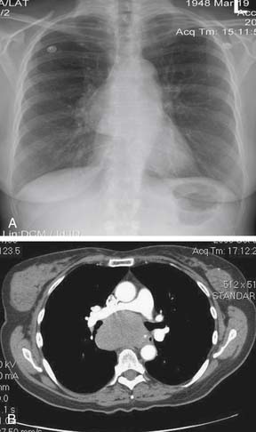
Computed tomography (CT), often with intravenous or oral contrast, is indicated when mediastinal pathology is suspected on the basis of the chest film (see Fig 42-1B) or clinically. High-resolution spiral CT, with cross-sectional imaging of structures at intervals as narrow as 1 mm, is the radiologic modality of choice for imaging the middle mediastinum. All structures located in the middle mediastinum can be seen by CT. In addition, the relationship of these structures to other nearby structures can be delineated. Three-dimensional CT images can be reconstructed using computerized programs without the need for additional radiation. These reconstructions can be particularly useful for detailed assessment of structures, such as the trachea, located in the middle mediastinum.


Magnetic resonance imaging (MRI) may at times provide additional information in the assessment of middle mediastinal pathology. Advantages of MRI include its ability to differentiate between vascular, solid, and fluid elements in a given mass. However, MRI is most likely to be useful in the evaluation of posterior mediastinal masses (e.g., paravertebral tumors).


Positron emission tomography (PET) is a valuable tool for evaluating mediastinal pathology, especially mediastinal lymph node involvement in neoplasms (e.g., lung cancer). The degree of uptake of fluorodeoxyglucose (FDG) labeled with radioactive fluoride (18F) is a surrogate marker of the degree of metabolic activity in a cell. However, PET is limited, because it is difficult to distinguish between increased metabolic activity caused by tumor and that caused by inflammation. Furthermore, as PET does not provide an anatomic scan, the precise location of the increased metabolic activity may not be discernable. However, fusion of PET with CT (CT-PET) has allowed a more precise assessment of anatomy.1


Conventional ultrasonography is of limited value in the evaluation of middle mediastinal pathology, although it may help in determining whether a mass is cystic or solid. However, transesophageal endoscopic ultrasonography (EUS) has emerged as a valuable tool for the evaluation of certain mediastinal pathology, especially lymph nodes.2 Other modalities, such as leukocyte scintigraphy, lymphoscintigraphy, and metaiodobenzylguanidine (MIBG) scanning, have limited and very specific indications in the evaluation of pathology in the middle mediastinum.

< div class='tao-gold-member'>

Only gold members can continue reading. Log In or Register to continue

Stay updated, free articles. Join our Telegram channel

Jul 30, 2016 | Posted by in CARDIAC SURGERY | Comments Off on The Middle Mediastinum

Full access? Get Clinical Tree

Get Clinical Tree app for offline access