The Mediastinum, Its Compartments, and the Mediastinal Lymph Nodes
Thomas W. Shields
The mediastinum is defined as the thoracic space that lies between the two pleural cavities. It extends from the thoracic inlet cephalad to the superior surface of the diaphragm caudad. It is bounded by the undersurface of the sternum ventrally and the anterior longitudinal spinal ligament dorsally. The paravertebral areas (the costovertebral sulci) situated bilaterally are not truly within the mediastinum, but lesions arising within these regions are classically defined in medical literature as mediastinal in origin.
Subdivisions of the Mediastinum
Numerous radiographic and surgical subdivisions of the mediastinum have been used in the literature. The more commonly used anatomic subdivisions are the superior, anterior, middle, and posterior mediastinal areas. The boundaries and locations of these subdivisions are defined differently by the many authors who have written on the subject. As a consequence, confusion abounds in the radiologic, pediatric, and surgical literature about these divisions. The same organ or structure may be located in two or more regions as it passes from the thoracic inlet to the diaphragm. More important, these commonly used divisions place the trachea and the esophagus, both derived from the foregut, in different compartments, which is inappro- priate.
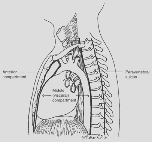
In 1972, the author4 suggested that a simple anteroposterior division of the space be used (Fig. 162-1). This schema consists of an anterior compartment, a visceral compartment, and the paraventral sulci bilaterally. At the thoracic inlet, the visceral compartment occupies the entire space anterior to the vertebral column, and the two paravertebral sulci are posterior and lateral to the anterior surface of the spinal column. The anterior compartment (the prevascular space) is limited superiorly by the innominate vessels and does not communicate directly with the thoracic inlet (Figs. 162-2 and 162-3), although the space may be entered through the inlet by the appropriate surgical techniques (see Chapter 169). Each compartment extends inferiorly to the diaphragm and is limited laterally by the mediastinal surface of the respective parietal pleura. The anterior compartment is bounded anteriorly by the undersurface of the sternum inferior to the innominate vessels and posteriorly by an imaginary line formed by the anterior surfaces of the great vessels and pericardium. The visceral compartment (also referred to as the postvascular space, the middle mediastinum, or the central space) extends from the posterior surface of the superior portion of the sternum above the innominate vessels and the posterior limit of the anterior compartment below these vessels to the ventral surface of the vertebral column. Thus, as noted, the visceral compartment occupies the thoracic inlet. Often, it is convenient to divide the superior portion of the visceral compartment at or just below the thoracic inlet into
pretracheal and retrotracheal areas. The paravertebral sulci (costovertebral regions) (Fig. 162-4), as noted, are not truly mediastinal in location but are potential spaces along each side of the vertebral column and adjacent proximal portions of the ribs.
pretracheal and retrotracheal areas. The paravertebral sulci (costovertebral regions) (Fig. 162-4), as noted, are not truly mediastinal in location but are potential spaces along each side of the vertebral column and adjacent proximal portions of the ribs.
 Figure 162-1. Schematic illustration of the mediastinal subdivisions and the paravertebral sulcus as seen from the left. Note that the anterior compartment does not extend to the thoracic inlet but is limited superiorly by the brachiocephalic (innominate) vessels.
Stay updated, free articles. Join our Telegram channel
Full access? Get Clinical Tree
 Get Clinical Tree app for offline access

|