The Functioning Normal Heart
Coordination of the Heartbeat, the Valves, and the Flow of Blood
To understand an engine, it isn’t enough to learn about its parts. You have to turn on the machine and watch it function. How is the heartbeat coordinated in various chambers? What keeps the blood from going backward instead of forward? How is the heartbeat timed so that each chamber is ready to receive the inrush of blood at the right split second? Watch a sample of blood move through the heart-lung machine and see how everything is coordinated.
Systole
The ventricles contract and the pressure inside them rises. When the pressure inside the ventricles is higher than the pressure inside the atria, the atrioventricular (AV) valves are slammed shut, just as if the pressure of your hand was closing a door. In other words, the mitral and tricuspid valves are forced shut early in ventricular systole. The inrush of blood from the veins fills the atria against these closed valves.
The same rise in pressure forces the outlet valves open, so that blood can flow out of the ventricle into the pulmonary artery and the aorta. The aortic and pulmonic valves are forced open early in systole (Fig. 5-1).
Diastole
The ventricles relax and the pressure in them falls. When it falls below the pressure in the atria, the AV valves swing open and blood rushes into the ventricles. The mitral and tricuspid valves are forced open early in diastole.
The same fall in pressure drops the pressure in the ventricles below the pressure in the great vessels (the aorta and the pulmonary artery). The higher pressure in
the great vessels forces the outlet valves shut, holding the blood in the lungs and in the whole body.
the great vessels forces the outlet valves shut, holding the blood in the lungs and in the whole body.
The aortic and pulmonic valves are forced shut early in diastole (Fig. 5-2).
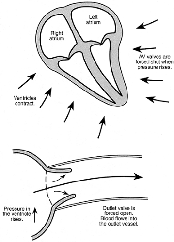 FIGURE 5-1 The events of systole. |
The Electric Circuit That Drives the Heart
The muscles that move your body can’t function by themselves: they can only contract when they are stimulated by a nerve impulse. This impulse is actually a wave of electric energy that starts in the brain and travels down a nerve to the muscles involved in a particular motion. If the nerve is cut or if the brain tissue is destroyed, the muscles will be paralyzed (Fig. 5-3).
The heart is different. It has its own self-contained “nerve” system to stimulate it to beat. The cells that make up this system are really heart muscle cells, specially adapted, but they function as nerve cells.
The electric energy that stimulates the heart muscle begins in a small bundle of tissue less than 0.5 inch long located near the top of the right atrium. This very important
structure is called the sinoatrial node, often abbreviated as the SA node. The sinoatrial node is the source of the electric energy that makes the heart beat: it builds up a definite potential and then discharges, sending a wave of electric energy spreading across the atria, like a ripple across a pond. When muscle is stimulated by an electric impulse, it contracts (you probably remember the way your hand jerked when you touched a live light socket as a child). The cells of the atrial walls transmit the activating impulse from cell to cell. This meshwork of cells is called the atrial syncytium (Fig. 5-4).
structure is called the sinoatrial node, often abbreviated as the SA node. The sinoatrial node is the source of the electric energy that makes the heart beat: it builds up a definite potential and then discharges, sending a wave of electric energy spreading across the atria, like a ripple across a pond. When muscle is stimulated by an electric impulse, it contracts (you probably remember the way your hand jerked when you touched a live light socket as a child). The cells of the atrial walls transmit the activating impulse from cell to cell. This meshwork of cells is called the atrial syncytium (Fig. 5-4).
 FIGURE 5-2 The events of diastole. |
There is a bundle of conducting tissue leading out of the atria, like a small canal leading out of a pond. This bundle connects the atria and the ventricles and is logically named the atrioventricular or AV node. The cells in the atrioventricular node are specialized: they conduct the activating current of the heart just as an electric wire might conduct current from a battery to a lightbulb. The lower end of the AV node is called the bundle of His—it’s a little different in structure from the upper part. The bottom of the bundle of His divides into two branches, one to each ventricle. Logically, these are called the left and right bundle branches. The bundle branches divide into progressively smaller twigs, exactly like the branches of a tree. They end in small fibers called the Purkinje fibers. These tiny elements actually transmit the electric charge to the heart muscle (Fig. 5-5).
Stay updated, free articles. Join our Telegram channel

Full access? Get Clinical Tree


