Structure and Function of the Normal Heart
Before you begin to learn about heart disease, you must learn how the normal heart is constructed and how it functions. This is easier than you might think, because the heart is a surprisingly simple organ. An hour’s easy reading will give you all the information you need to begin.
The Chambers of the Heart and their Connections
The heart is a hollow organ divided into four chambers, two on the top and two on the bottom (Fig. 1-1). Study this simple diagram until you know it as well as your own name: it’s basic to everything else in the book.
The top two chambers are thin-walled structures that act primarily as holding chambers for the blood. They are called atria. This is the plural of the Latin word atrium, meaning “anteroom” or “porch,” and, in fact, these chambers do act as entryways to the great chambers below. The ventricles are large, thick-walled chambers that do the real work of pumping the blood. (This name comes from the Latin ventriculum, meaning a “cavity” or “pouch.”)
Look again at Figure 1-1 and note the wall, or septum, that divides the left atrium from the right atrium and the left ventricle from the right ventricle. This wall of tissue is much like the septum in your nose that separates the two nostrils. The important thing to remember about the heart’s septum is that it is absolutely watertight, or, more properly, “bloodtight.” Normally, no blood can pass through this septum from one side to the other. (It took the human race about 4,000 years to discover this simple fact. The ancient Greeks and Romans were convinced that blood somehow oozed through the septum from one side to the other. It doesn’t.)
Physicians commonly refer to the right atrium and right ventricle together as the right heart and to the left atrium and left ventricle as the left heart.
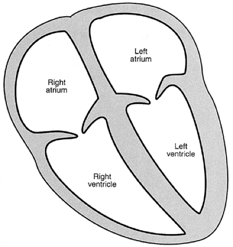 FIGURE 1-1 The chambers of the heart. |
The Motion of the Blood Through the Heart
The function of the heart can be described as a simple pump that forces blood forward by squeezing, in exactly the way that a bulb syringe forces out fluid when it’s compressed.
The alert reader will at once ask, “If the blood doesn’t flow from one side of the heart to the other through the septum, how does it ever move forward?” The answer to that question eluded philosophers and scientists until the English medical doctor William Harvey, in the early seventeenth century, discovered the simple circuit that is the basis of all modern cardiology.
Stay updated, free articles. Join our Telegram channel

Full access? Get Clinical Tree


