The Diabetic Foot

Chapter 59 The Diabetic Foot


Diabetes is a growing epidemic in the United States and around the world. The 2011 National Diabetes Fact Sheet estimates that 26 million children and adults in the United States have diabetes, representing 8% of the U.S. population. An additional 79 million have prediabetes but nevertheless may be susceptible to the full range of its complications. Diabetes complications, which include cardiac disease, stroke, hypertension, blindness, renal disease, neuropathy, and amputation, consume more than 30% of U.S. health care expenditures.1


Many are surprised to learn that foot disorders, such as ulceration, infection, and gangrene, are the leading causes of hospitalization in patients with diabetes mellitus. Approximately 15% to 20% of persons with diabetes will be hospitalized with a foot complication at some time during their lifetime. Ulcerations are commonly painless and therefore perceived by the patient to be innocuous. Amputations are preceded by a foot ulcer in more than 85% of cases. The final steps on the pathway to major amputation are ischemia and severe infection. The effect of these concurrent factors can make major emergency amputation a life-saving procedure.


More than 80% of nontraumatic lower limb amputations occur in patients with diabetes. Current estimates place the number of amputations in the United States at 80,000 per year. Direct and indirect costs in medical care, hospitalizations, and disability accruing in the wake of the diabetic foot constitute a significant portion of the federal health care budget. This pandemic collectively represents a health care calamity and a crushing financial burden. These figures make it imperative that our society and our profession do more to achieve the twin linked goals of ulcer healing and amputation prevention.


Neuropathy


Peripheral neuropathy is one of the most common long-term complications of diabetes, affecting up to 50% of patients by the tenth year of the diagnosis. It typically manifests as a mixture of sensory, motor, and autonomic involvement. The pathogenesis of diabetic neuropathy results from metabolic and vascular disorders that affect nerve blood flow and vascular endothelial function.2


Patients are usually divided into two groups: painful neuropathy or painless neuropathy. Painful neuropathy occurs in 15% of cases, with patients complaining of burning, lancinating pain, or allodynia. Various treatments exist to treat the symptoms of painful neuropathy, including antidepressants and anticonvulsants, which modulate neurotransmitters in the central nervous system. Other treatments for pain including surgical decompression, infrared light therapy, or vitamins are unproven. Painful neuropathy usually becomes painless in 2 to 3 years; therefore treatments for pain can be reduced or even eliminated at that time. Painless neuropathy or sensory neuropathy would seem to be preferable to the painful variety; however, it is this type of neuropathy that leads to the ulcerative complications in the diabetic foot. There are no accepted treatments to reverse sensory neuropathy in diabetes, and clinicians should not be lured into marketing traps by vitamin companies or surgical decompression enthusiasts who are without credible evidence to support their claims. A minority of patients have a syndrome with characteristics that are both painful and painless. In addition, ischemic rest pain symptoms induced by lower extremity arterial insufficiency in diabetes can often be difficult to distinguish from neuropathy.


A neurologic examination should be done to evaluate sensory and motor function in all patients with diabetes. Loss of protective sensation (LOPS) is a more severe form of diabetic neuropathy that increases the risk for ulceration. It is most readily revealed by applying pressure on selected dorsal and plantar areas of the foot with a deformable Semmes-Weinstein monofilament (SWMF) (Figure 59-1).2


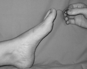
If the patient is unable to feel 10 g of pressure exerted by a SWMF, they have met the criteria for LOPS. Patients should be screened annually for neuropathy. The nerve function profile of Boulton and colleagues provides a useful and reproducible baseline that has been validated as predictive of ulceration.3,4


Because diabetic neuropathy affects all three divisions of the peripheral nervous system, the motor signs are sometimes subclinical but can lead to disabling disease progression manifested by muscular atrophy, fasciculation, and weakness. Intrinsic muscle weakness of the foot, once it becomes pronounced, leads to digital contractures and secondary metatarsal-phalangeal instability. The extrinsic muscles gain mechanical advantage over the intrinsic, which results in an unstable foot, prominent metatarsal heads, plantar callus formation, and ulceration. The sensory distribution is predominately of the feet and to a lesser extent the hands. Rather than following specific nerve trunks, the pattern is one of a stocking or glove. Motor neuropathy produces a pronounced clinical finding that is obvious on even cursory examination while the medical history is obtained; interosseus muscle wasting of the hands is evident, sometimes dramatically so.


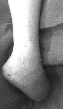
Autonomic neuropathy interrupts control of arterial and capillary flow to the feet and tissues, resulting in decreased sudomotor function and dry feet, anhidrosis, noncompliant skin, and, ultimately, fissuring and painful cracking of the skin. Deep heel cracks and fissures are a common manifestation of autonomic neuropathy. This process results in an “auto-sympathectomy,” which explains why surgical ablation of the lumbar sympathetic trunk provides no benefit for diabetic ulceration. The stage is thus set for bacterial penetration and clinical infection. Autonomic control of vascular smooth muscle can also be affected, which leads to hyperemic states and possibly contributes to the development of Charcot destruction. The importance of neuropathy in diabetic foot ulceration is best encapsulated by the words of Dr. Paul Brand, considered “the patron saint of the diabetic foot.”4a He wisely observed: “If the patient walks into your clinic with a diabetic foot ulcer and does not limp, he has neuropathy. For that reason alone the doctor should always observe the patient as he/she walks.”


Ulceration


Evaluation


A review of the clinical examination reports reveals that doctors at all levels of training who see patients for foot ulceration often fail to describe the appearance, size, and location of the lesion, or the state of infection in the presenting complaint. Patients should remove their socks and shoes during routine clinical evaluations.


The size, depth, and location of ulcers should be measured at the first examination and serially documented thereafter to track progression. The use of photographs to document ulcer size and location is recommended. Clinical findings of infection, such as the presence of malodor, exudate, and erythema, are obvious, but in diabetics, clinical manifestations can also be subtle. Clinical signs may help elicit the bacterial pathogen. Malodorous drainage suggests an anaerobic infection; creamy, yellow pus indicates a Staphylococcus species infection; and thin, watery drainage on a green base may herald infection with Pseudomonas species. Probing the depth of the ulceration with a metal probe can reveal involvement of tendon sheaths, open fascial planes, or abscess formation. Because of LOPS, this procedure rarely elicits patient complaint.


The evaluation should also identify common deformities and biomechanical abnormalities that contribute to the ulcer. For instance, ill-fitting footwear can cause ulcers to develop on the dorsum of toes, interdigitally, or on the lateral aspect of the foot. Ulcerations located on the dorsal aspect of toes may be related to contracted digits in a shallow toe box. Ankle equinus contracture, defined as ankle dorsiflexion of less than 10 degrees, causes increased pressure of the plantar forefoot and midfoot, leading to calluses and ulceration. Patients with amputations or shortened foot are susceptible to ankle equinus because the ankle flexors overpower the extensors (Figure 59-2).5


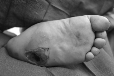
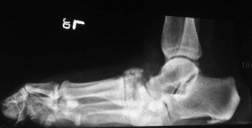
The Charcot foot rocker bottom deformity increases pressure at the apex of the deformity, usually at the midfoot, leading to mid-foot collapse and to the characteristic ulcer (Figures 59-3 and 59-4).


The plantar ulcerations that arise with increased ankle flexion and plantar pressure are not usually amenable to correction with footwear; the Achilles tendon is best dealt with by tendo-Achilles lengthening (TAL) or operative lengthening of the gastrocnemius muscle.6


Ulcer Classification


The established systems to stratify the severity of foot ulceration are based on the size of ulceration (area and volume), location, tissue loss, presence of infection, and the anatomic and hemodynamic state of the arterial circulation. Three diabetic ulcer classification systems are widely used: the University of Texas (UT) classification, PEDIS, and Wagner grading.7,8 The UT and PEDIS classifications were developed specifically for the diabetic foot. Wagner grades were first described for the foot known or presumed to be dysvascular but are now applied to all etiologies of lower extremity ulcerations.9 The UT system appears to be a better predictor of ultimate wound outcome, is often used in wound centers, and has been validated. The Wagner classification is based on the depth of penetration, the presence of osteomyelitis, or the extent of gangrene. A major shortcoming of Wagner grading is that the presence of ischemia is not specified in the consideration of the earlier stages. Because most ulcers fall into either grade I or II, the Wagner system inadequately differentiates the etiologic factors that underlie the majority of lesions. Finally, there is the (P)erfusion, (E)xtent, (D)epth, (I)nfection, (S)ensation or PEDIS classification developed by the International Consensus on the Diabetic Foot, which was devised initially as a research tool because of its relative complexity (Table 59-1).


TABLE 59-1 Table of Ulcer Classification Systems


System Classifications
Wagner
University of Texas diabetic foot wound classification system
PEDIS


Ulcer stratification continues to loom as a major problem. Without an effective system around which investigators can find agreement and consensus, our ability to compare patient populations, understanding disease severity, and improving effectiveness of treatment will remain an unattainable goal.


Charcot Foot


Charcot neuroarthropathy was first described in tabes dorsalis in syphilis. It is now most commonly associated with diabetes but could occur as a result of any neuropathy.10 The International Task Force on the Charcot Foot stated the condition is primarily a disease of unopposed inflammation in the foot, leading to increased vascularity and bone resorption. It is a chronic and most often progressive disease of bone and joints. Common features include joint dislocation, pathologic fractures, and severe destruction of the pedal architecture, resulting in debilitating deformity, increased risk of ulceration, and increased risk for major amputation from complications of ulceration. It can lead to disability and premature retirement. The task force recommended classifying Charcot foot as active or inactive, depending on whether inflammation was present.11


The most important factor in early detection and the potential to alter the outcome of patients with Charcot foot comes from retaining a high clinical suspicion in patients with the important risk factors. It can be precipitated by seemingly minor trauma. The clinical signs most associated with the disease are an erythematous, hot, and edematous foot. When these clinical findings predominate, it is easy to understand why Charcot can be mistaken for, and is treated as, a deep infection. Approximately 50% of patients have pain with their Charcot foot, which complicates its diagnosis because it is a syndrome caused by neuropathy. The diagnosis is often missed and thus delayed. The characteristic rocker bottom deformity is a late sign of the disease.12


Plain radiographs meant to find subtle changes or subluxations should be the initial diagnostic procedure. Magnetic resonance imaging (MRI) or bone scintigraphy can confirm the diagnosis in some cases, but usually clinical suspicion is enough to make the presumptive diagnosis and begin treatment because the consequences of undertreatment or no treatment can be devastating.


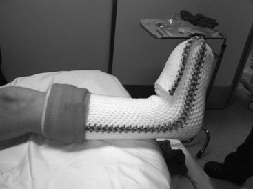
There is consensus that the cornerstone of management of acute Charcot foot is early immobilization and offloading. Judicious use of a total contact cast (TCC) or removable cast walker, crutches, walkers, knee walkers, wheelchairs, or bed rest may be appropriate in this setting (Figure 59-5).13,14


Immobilization for 4 to 6 months or more may be necessary until inflammation of the extremity becomes quiescent. Skin temperatures are generally 8° F to 10° F (−13.3° C to −12.2° C) hotter on the affected side. Skin temperatures are used to determine when the inflammation finally becomes inactive. In general, a difference of 4° F or less is considered normal. Adjuvant use of bisphosphonates or low-intensity ultrasound bone stimulation has proved ineffective. Prolonged bracing with Charcot Restraint Orthotic Walkers (CROWs) may be necessary for long-term protection.


Ulcer Management


After adequate ulcer evaluation, wounds are managed with nonsurgical or surgical strategies.


Nonsurgical


Nonsurgical treatments include topical wound agents, dressings, and hyperbaric oxygen therapy (HBOT). Topical wound agents and dressings historically served as methods for infection control. Today, a myriad of dressing options is available to assist with moisture, exudate, and bacterial control, as well as with delivery of growth factors. Bioengineered tissues containing living fibroblasts can be applied to a well-vascularized, uninfected ulcer and improve granulation tissue and lead to wound closure. Once infection has been eradicated, potentially harmful topical agents, like povidone iodine, should be diluted or avoided and wounds should remain moist to limit tissue desiccation.


Offloading is an essential complement to all forms of therapy. Offloading assists with ulcer healing by decreasing mechanical stress, shear, and trauma. Patient compliance, however, is often problematic because patients in many cases do not have pain and are not aware that repetitive trauma is occurring. Although the TCC has been reported as the gold standard for offloading, it is not widely used given the difficulty and time required for application and the potential complications. There are other alternative offloading devices and strategies such as non–weight-bearing with crutches, walkers, or wheelchairs, or bed rest. However, attempting to keep patients at least partially weight-bearing with removable cast walkers, knee walkers, half shoes, and prescription accommodative shoes is the goal.


The rationale for HBOT for diabetic ulceration is presumed cellular proliferation and angiogenesis. The patient is placed in a single or multiplace compression chamber set at 2 to 3 atm of pressure and inhales 100% oxygen, thereby dramatically increasing the oxygen partial pressures in tissues. Under normal atmospheric pressure, erythrocyte hemoglobin is essentially saturated while little oxygen is transported by blood plasma. Oxygen transport by plasma is significantly increased using HBOT. Reported effects of increased oxygenation include enhancing antibacterial effects, increasing fibroblast activation, upregulation of growth factors, promotion of collagen synthesis, mobilization of stem/progenitor cells from the bone marrow, and angiogenesis. Specific indications for HBOT include wounds with deep soft tissue infection, necrotizing infection, compromised skin grafts and flaps, and refractory osteomyelitis. Topical HBOT has not been shown to be of any clinical benefit.


Surgical


The diabetic foot has several surgical indications. When infection or gangrene is present, urgent operative therapy should be considered to remove infected or devitalized tissue. If a chronic wound fails to respond to therapy after 4 to 6 weeks, surgery to achieve wound closure may need to be considered.


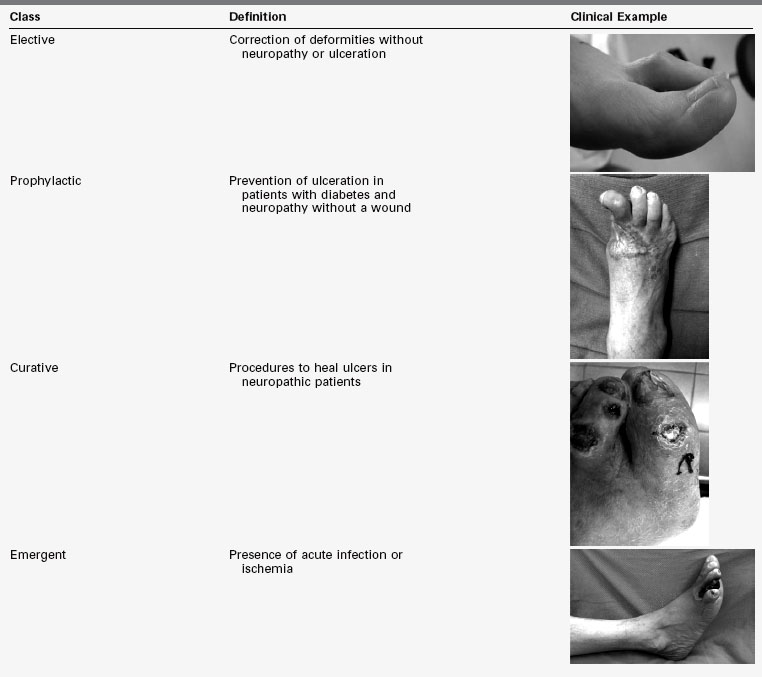
Diabetic foot surgical interventions fall into one of four classes: elective, prophylactic, curative, and emergent. Elective surgery is composed of procedures for deformities in the absence of neuropathy or ulcerations. Prophylactic surgery includes procedures for the prevention of ulceration in patients with diabetes and neuropathy, but without a wound. Curative procedures are used to heal ulcers in neuropathic diabetic patients. Emergent procedures are performed in the setting of acute infection or ischemia. The risk of postoperative infection increases as the classes progress from elective to emergent (Table 59-2).15


TABLE 59-2 Classes of Foot Surgery With Clinical Examples


image

Surgical debridement is advisable for diabetic foot wounds. It can be performed by scalpel, curette, or hydrosurgery using the Versajet (Smith and Nephew, London, U.K.). After debridement, a large soft tissue defect can remain. The use of negative pressure wound therapy (NPWT) leads to granulation of wounds with large soft tissue defects while controlling exudate and infection. NPWT works by causing microdeformational forces on the cells that stimulate division and replication. The goal of NPWT is not necessarily to heal wounds but to provide a granular wound bed level with the surrounding tissue. At this point, techniques to close the wound should be used.


When diabetic ulcerations are located on the foot and ankle over bony prominences where there is limited mobility of skin or lack of available tissue, and the surface is involved in a weight-bearing location, wound coverage can be challenging. Reconstruction should cover defects with tissues similar to lost ones while minimizing donor site morbidity. Methods include bioengineered living tissue and acellular matrices, skin grafts, local skin flaps, fasciocutaneous flaps, and muscle flaps. Acellular matrices often serve as a temporary scaffold upon which granulation tissue forms. This tissue prepares the wound for definitive closure with autologous skin grafting or bioengineered living tissue. Often skin grafts and flaps are required because of extensive loss of skin and soft tissue. Excessive skin tension can occur with primary closure, which can lead to skin necrosis and a dehisced wound; these complications can be avoided with meshed split thickness skin graft (STSG) or by using NPWT over incisions. STSG should be avoided whenever possible over weight-bearing areas because plantar skin must withstand the mechanical stress of weight-bearing and footwear. NPWT serves not only as a bolster over STSG or acellular matrices but also controls exudation and accelerates graft incorporation. NPWT is typically used over grafts continuously for 3 to 5 days and then discontinued.


Below-the-ankle amputations in diabetic patients are considered successful outcomes if they result in capable, balanced, plantigrade walking on a foot that is capable of limiting the formation of ulcers. Partial foot amputations including digit and partial ray amputation are often the end result of debridement initially performed to control infection. Secondary closure may be the final step in a series of staged procedures. In the presence of an acute infection, an open amputation achieves drainage, debrides necrotic tissue, and allows for infection to be treated with antibiotics before definitive closure or amputation revision. Definitive closure of the wound is delayed until vascularity is optimized. The preferred location of amputation level is the lowest level that would heal with the most functional remaining foot.


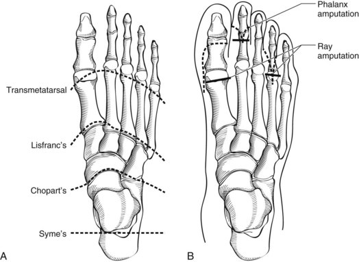
Forefoot amputations are desirable because they preserve length, which preserves maximal mobility and function; the transmetatarsal amputation (TMA) is a case in point. More proximal partial foot amputations such as at the Lisfranc joint or Chopart amputation at the talonavicular and calcaneocuboid joints require adjunctive procedures to maintain a balanced foot (Figures 59-6 and 59-7).


< div class='tao-gold-member'>

Stay updated, free articles. Join our Telegram channel

Jul 1, 2016 | Posted by in CARDIOLOGY | Comments Off on The Diabetic Foot

Full access? Get Clinical Tree

Get Clinical Tree app for offline access