Technical Aspects of Eversion Carotid Endarterectomy for Atherosclerotic Disease


Technical Aspects of Eversion Carotid Endarterectomy for Atherosclerotic Disease


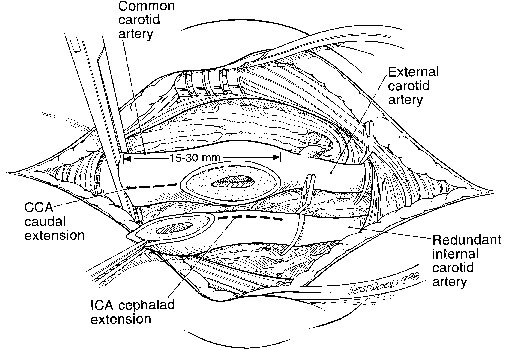
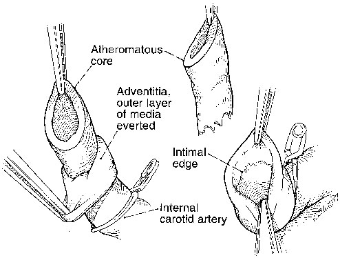
Dhiraj M. Shah, R. Clement Darling, III, Benjamin B. Chang, Paul B. Kreienberg, Philip S.K. Paty, Kathleen J. Ozsvath, Sean P. Roddy and Manish Mehta


All methods of arterial closure following a standard carotid endarterectomy (CEA) are associated with appreciable rates of persistent or recurrent internal carotid artery (ICA) stenoses, of which some are related to technical issues in closing the artery. Eversion endarterectomy of the ICA is an alternative technique that had its origins in the United States and recent resurgence in Europe. The eversion technique, as currently conceived, involves complete transection of the ICA at its origin from the carotid bifurcation. This procedure should be distinguished from the eversion endarterectomy involving division of the distal common carotid artery (CCA) as described by DeBakey and colleagues in 1959. The latter method is only of historical interest, because lengthy disease of the ICA is difficult to treat with this method.


Current eversion endarterectomy facilitates the removal of plaque higher up in the ICA. More importantly, this method appears to improve upon standard endarterectomy in two and possibly three ways. First, because the endpoint is fully visualized and generally not sutured, it is easier to detect intimal flaps. Second, and most clearly, the reanastomosis of the ICA to the bulb is technically simple and obviates the risk of narrowing a primary closure of the ICA during conventional endarterectomy or the need for patching. Third, this method has been associated with a decrease in later restenosis.


Methods


Surgeons adopting eversion endarterectomy need not change much of their conventional CEA technique. Initial dissection and isolation of the carotid artery are identical. However, after the artery is exposed, it is important to circumferentially isolate the ICA and the bulb. The need for more extensive dissection is a common criticism of this technique, but in reality it has not produced an increase in complications, most particularly, an injury of the vagus nerve. In addition, if desired, this dissection may be completed after cross-clamping, but this increases the cross-clamp time unnecessarily. After the carotid arteries are isolated, heparin sodium is administered and the arteries are clamped.


At this time, the ICA is transected obliquely, with the line of division running from the crotch of the carotid to a point more proximal on the lateral (internal) side of the CCA. The specific angle of division is not critical, but a 10- to 15-mm opening should be left in the CCA. Doing so aids visualization of disease in the bulb and facilitates reanastomosis of the arteries. After division, the ICA almost always is redundant. Because of this, it may be spatulated, further increasing the diameter of the eventual suture line. If this is done, the common carotid arteriotomy is extended caudad a similar amount (Figure 1).


Endarterectomy of the ICA is begun by circumferentially elevating the plaque from the arterial wall. It is important to remove both the intima and media, and if done, the adventitia may be grasped with two fine forceps while the assistant holds the plaque. The adventitia is then pulled or rolled like a sock until the end of the plaque is reached (Figure 2). If the endarterectomy plane is too shallow and appreciable media remains, the artery will be too stiff to easily evert. At this time, the specimen usually spontaneously divides at the endpoint. If not, the plaque is sharply divided at its termination. This process usually requires less than 30 seconds.


At this point, the assistant holds the luminal side of the adventitia as near as possible to the endpoint. This task can require the surgeon to move the ICA clamp more cephalad. The operator may then inspect the entire circumference of the endpoint, removing loose fragments and making sure the distal intima is adherent. It is critically important for the operator to see the entire endpoint clearly at this time. If a loose flap is found, it can usually be removed. Rarely, tacking sutures may be employed, and these are best performed using double-armed fine sutures inserted from the luminal side and tied externally.


After the endpoint is secured, the artery is unrolled and the interior is inspected for loose bodies. Irrigation with heparinized saline into the lumen allows any loose fragments to float away from the wall to aid in their removal. Any persistent stenosis or flap is then corrected. This is the most critical point of the operation and should be done carefully.


After the ICA endarterectomy is completed, the distal CCA and the external carotid artery (ECA) are examined. If there is no significant disease, the arteries may be reanastomosed. More often an extended endarterectomy is performed. The plaque is elevated in the bulb and carried up the ECA and proximally into the CCA as dictated by the extent of the disease. Division of the plaque in its midportion allows the surgeon to deal with each artery separately.


Endarterectomy of the ECA is performed by standard methods. Endarterectomy of the CCA may also be performed with a combination of direct elevation of exposed plaque and proximal eversion of more extensive plaque. Rarely, if the CCA plaque extends very proximally, the arteriotomy in the distal CCA can be extended inferiorly. The CCA is then primarily reapproximated or the ICA is opened further, to encompass the expanded CCA arteriotomy.


After the endarterectomy is completed, the ICA is reapproximated to the CCA. Because the original line of incision was oblique and both arteries were further opened, the anastomosis is usually 10 to 30 mm in length. Thus closure of the artery is much easier with the eversion technique, and there is little chance that the arteries will be narrowed during closure. A continuous suture of 6–0 polypropylene is started at the most cephalad portion of the internal carotid arteriotomy. Because the walls of the arteries are now redundant, fairly large bites may be taken with the suture with little fear of causing a stenosis. Following completion of the anastomosis, antegrade blood flow is reinstituted in the usual manner.

Stay updated, free articles. Join our Telegram channel

Aug 25, 2016 | Posted by in CARDIOLOGY | Comments Off on Technical Aspects of Eversion Carotid Endarterectomy for Atherosclerotic Disease

Full access? Get Clinical Tree

Get Clinical Tree app for offline access