Management of Acute Limb Ischemia Complicating Aortic Reconstruction


Management of Acute Limb Ischemia Complicating Aortic Reconstruction


Timothy Baxter and Jason MacTaggart


Scope of the Problem


Historical reports documented acute limb ischemia in up to 25% of patients undergoing abdominal aortic surgery. With refinement of operative technique and the use of local and systemic anticoagulation, the incidence of this complication has decreased significantly, but it has not been eliminated. Data from the randomized aneurysm trials and large retrospective database analyses report perioperative thromboembolic complication rates between 1% and 2% and amputation rates significantly less than 1% for both open and endovascular reconstruction techniques. Though patients with combined aneurysmal and occlusive disease patterns seem to have the highest risk, most reports demonstrate no major differences in occurrence between patients operated on for aneurysmal disease compared to occlusive disease.


Etiology


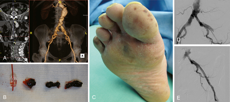
There are a number of causes of acute limb ischemia associated with aortic surgery and they can be broadly classified as embolic or thrombotic (Figure 1).


Embolic subcategories include macroemboli and microemboli (see Figure 1B and C). Macroembolic sources found in diseased aortas, such as laminated or friable thrombus (see Figure 1A), can be dislodged during dissection, clamping, or endovascular manipulations and can result in obstruction of the major named arteries supplying the lower extremity. In the absence of adequate anticoagulation, thrombus can quickly develop in the obstructed artery. In cases of microembolism, the major arteries of the leg remain patent while small fragments of thrombus or debris lodge in the smaller vessels and microcirculation of the lower limb. Atheroembolism, the disruption and release of atherosclerotic plaque contents, is one type of microembolic syndrome that can produce the classic trash foot, but these microparticles can also lodge in the muscles of the legs and buttocks.


Thrombotic etiologies relate primarily to inflow, conduit, and outflow problems. Poor inflow can cause thrombosis of the native artery or graft and is commonly caused by clamp injury, unappreciated proximal stenotic disease, and the use of large occlusive sheaths. Defects in the conduit, such as twisted, kinked, or compressed graft or stent graft limbs can also result in thrombotic complications. Disadvantaged outflow, such as anastomotic defects, arterial wall dissections, and flaps (see Figure 1D and E) or heavily diseased distal vasculature, can give rise to abnormal flow, leading to thrombus formation and acute limb ischemia. Lastly, systemic conditions, such as profound hypotension and hypercoagulability, can also cause thrombosis of the reconstruction or downstream arteries.


Diagnosis


The diagnosis of acute limb ischemia following aortic surgery relies principally on the physical examination. Lower extremity pain, paresthesias, and paralysis may be present in patients who are awake and alert, but in the unconscious patient, these findings cannot be readily identified. Intraoperatively, iliac artery occlusion may be identified by the absence of backbleeding. After excluding retractor compression, a catheter is typically passed through the iliac artery in an attempt to retrieve thrombus or embolus. In a calcified and tortuous iliac artery, it may be safer and more effective to pass a red rubber catheter rather than a Fogarty balloon catheter. The red rubber catheter is then retrieved while suction is maintained. If backbleeding and flow cannot be established after reperfusion, consideration should be given to extending the graft to the femoral level or performing a femorofemoral bypass. The absence of a femoral pulse when one is anticipated typically necessitates exploration of the groin, open assessment of the common femoral artery, and selective thrombectomy.


After the procedure is completed and while maintaining a sterile operative field, the legs and feet can be inspected to identify most ischemic complications. The examination should include palpation of pulses and inspection of the legs and feet (see Figure 1C). The feet may be under the surgical drapes. Most importantly, knowledge of the preoperative lower extremity pulse and Doppler examinations is critical to compare with the postoperative examinations. Changes from baseline or failure to observe the anticipated improvement should be cause for concern and should prompt further investigation. Rarely, such as in cases of profunda femoris revascularization where gradual improvement is anticipated, the patient may be monitored closely without immediate intervention.


In some cases, noninvasive vascular studies are useful to help objectify one’s clinical impression and guide surgical decision making. Determination of the ankle–brachial index can provide an objective assessment of perfusion pressure and ischemic severity, which may be particularly important in an unresponsive patient with preexisting lower extremity occlusive disease. In addition, arterial duplex can be rapidly done at the bedside or in the operating room to directly visualize the arteries and to detect emboli, dissections, and other flow abnormalities for more precise diagnosis and therapy.


If the diagnosis or anatomic location of the problem is uncertain, conventional angiography performed in the operating room is the preferred imaging modality. With the proliferation of hybrid suites employing high-quality fixed imaging units immediately available within the operative field, there is little reason to avoid angiography if a problem is suspected. Computed tomography angiography (CTA) or magnetic resonance angiography (MRA) is occasionally useful in the immediate postoperative period once the patient has left the operating room, but these studies consume extra time, require larger contrast loads, and do not offer the option of definitive simultaneous intervention should a problem be detected.

Stay updated, free articles. Join our Telegram channel

Aug 25, 2016 | Posted by in CARDIOLOGY | Comments Off on Management of Acute Limb Ischemia Complicating Aortic Reconstruction

Full access? Get Clinical Tree

Get Clinical Tree app for offline access