Technical Aspects of Conventional Carotid Endarterectomy for Atherosclerotic Disease


Technical Aspects of Conventional Carotid Endarterectomy for Atherosclerotic Disease


Norman R. Hertzer


Carotid endarterectomy (CEA) is a conceptually simple but technically unforgiving surgical procedure. Although other factors like patient selection and preoperative antiplatelet therapy also play an important role, a good clinical outcome after CEA probably is related most directly to the technical perfection of the operation itself. Provided they are accurate, data from the Nationwide Inpatient Sample suggest that as many as 90% of the CEAs in the United States were done for asymptomatic carotid stenosis from 2005 to 2007. If for no other reason, therefore, the prevention of technical complications has never been more important. There is more than one way to perform CEA safely, but the following remarks describe the strategy the author adopted during almost 30 years at the Cleveland Clinic.


Surgical Technique


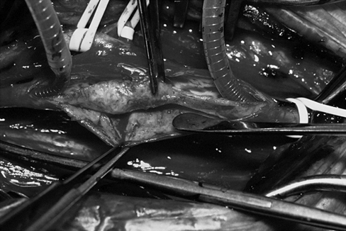
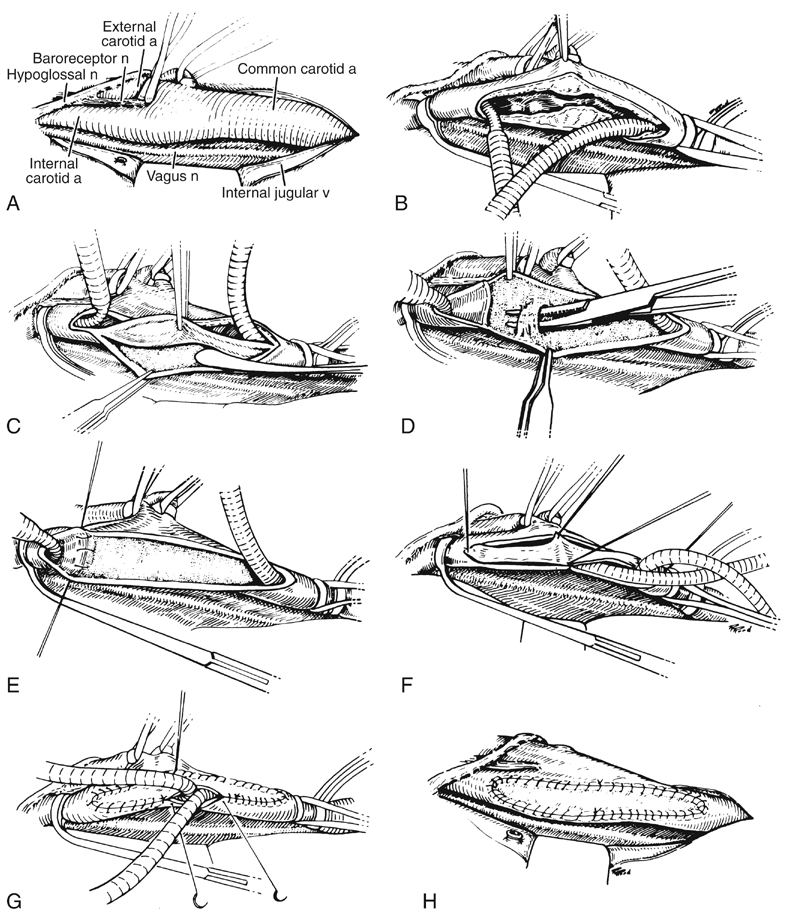
The author has previously reported a personal series of 2262 CEAs, nearly all being done under general anesthesia with routine carotid shunting and, during the last 20 years, patch angioplasty preferentially using the saphenous vein. The principal technical steps of the procedure became standardized (Figure 1).


After the patient is positioned on a shoulder roll, the neck, the upper chest, and the groin from which the greater saphenous vein is to be harvested are prepared and draped in a standard fashion. The cervical incision is made parallel and slightly anterior to the course of the sternocleidomastoid muscle, its length being determined by the level of the carotid bifurcation on preoperative imaging studies. If an especially high incision is necessary, it is angled posterior to the earlobe to avoid the parotid gland. The platysma layer is divided with the cutting mode of the electrocautery unit, and complete hemostasis is maintained throughout the operation, which generally is conducted within avascular planes. The internal jugular vein is reflected laterally with minimal exposure, ligating the common facial vein with a 5–0 suture before it is divided. The carotid sheath is opened longitudinally over the common carotid artery (CCA), being sure to avoid the vagus nerve if it happens to take an anomalous course anterior to the artery. In fact, the identification and preservation of several cranial nerves are cardinal features to the remainder of the dissection (see Figure 1).


The vagus nerve ordinarily lies within the carotid sheath posterior or slightly posterolateral to the CCA, may be closely adherent to the carotid bulb, and becomes nearly confluent with the hypoglossal nerve near the styloid process. To avoid at least temporary paralysis of the ipsilateral vocal cord, the vagus nerve should be protected from injury during the placement of self-retaining retractors, the application of a proximal arterial clamp, and the use of electrocautery. The hypoglossal nerve often is shrouded by small veins that never should be clamped and divided indiscriminately. This nerve probably is more likely to be injured by retraction than by full mobilization and, for this reason, structures tethering it in place—such as the artery and vein to the sternocleidomastoid muscle, the descending hypoglossal branch to the ansa cervicalis, and less commonly, the occipital artery—might require division to elevate the nerve for distal exposure of the internal carotid artery (ICA). The superior laryngeal nerve is located medial to the ICA, distributes branches that wrap around the superior thyroid artery, and can potentially be injured while controlling either of these two vessels. The glossopharyngeal nerve crosses the ICA near the base of the skull, often is filamentous and difficult to identify, and is best protected by maintaining dissection very close to the anterior surface of the artery. Finally, excessive or prolonged retraction at the upper aspect of the incision can cause at least temporary compression injuries laterally to the greater auricular nerve or medially to the marginal mandibular branch of the facial nerve.


It is well known that exposure should be obtained with minimal manipulation of the carotid bifurcation to avoid cerebral embolization. There should be no hesitancy in dividing the posterior belly of the digastric muscle superiorly, or the omohyoid muscle inferiorly, to facilitate atraumatic dissection and a subsequent arteriotomy that adequately encompasses the diseased segment of the carotid artery. Dissection near the baroreceptor nerve of Hering occasionally provokes bradycardia with associated hypotension, but this usually can be promptly controlled by the injection of 1 to 2 mL of local anesthetic into the carotid sinus at the notch of the bifurcation. A syringe containing 1% lidocaine should be available on the instrument table for this purpose.


Once the exposure has been completed and the patient has been systemically heparinized, traction is placed on the elastic vessel loops surrounding the external carotid and superior thyroid arteries. The proximal CCA then is controlled with an angled Hydrogrip clamp, after which a smaller clamp is applied to the distal ICA. The arteriotomy is begun with a No. 15 or No. 11 scalpel blade on the anterolateral aspect of the distal CCA. Using Potts scissors, the arteriotomy then is extended a sufficient distance proximally and distally to expose the full extent of the atherosclerotic plaque shown by preoperative imaging. Because of their eccentric calcification, some plaques promote a spiral arteriotomy through the internal carotid bulb unless care is taken to avoid it. Some thickening of the medial layer in the posterior wall of the CCA is not uncommon and can extend all the way to the innominate artery or the aortic arch. This generally should not be pursued, but further exposure of the CCA may be warranted if the lower end of an unexpectedly thick plaque can be palpated near the clavicle.


The distal end of a flexible loop shunt is then inserted into the ICA, retaining it with a small ring clamp. After all air is vented from the shunt, and the cul-de-sac of the CCA is filled with blood, the proximal end of the shunt is placed well into the CCA, temporarily preventing flow in the shunt by digital compression. After a proximal ring clamp has been positioned, the Hydrogrip clamp is removed by an assistant, and any potential debris is briefly flushed through the CCA around the shunt before the surgeon tightens the ring clamp and releases digital compression of the shunt. Another elastic vessel loop should be placed on slight traction around the CCA above the proximal ring clamp to prevent dislodgment of the shunt during the remainder of the procedure (see Figure 1). Any annoying backbleeding from the external carotid artery (ECA) usually can be controlled by applying a small hemostatic clip to an ascending pharyngeal branch hidden within the soft tissue of the carotid sinus.


The endarterectomy is begun using a Cannon blade or a Freer elevator and entering the alabaster subadventitial plane to excise all circular fibers of the media (Figures 1 and 2). The CCA endarterectomy is terminated about 1 cm above the proximal end of the arteriotomy. After the specimen is divided at this level, it can be elevated under full vision while the endarterectomy is continued upward into the carotid bulb. A plaque extending only a short distance into the ICA may be teased medially toward the origin of the ECA to achieve an adequate endpoint. More often, it is simpler to divide the plaque in the bulb so that the ICA and ECA endarterectomies can be done independently. Once the plaque has been divided and the elastic vessel loop has been loosened around the ECA, the ECA endarterectomy is performed with slight traction on the plaque to evert the artery until an appropriate endpoint has been attained. The ECA then is gently explored with the tips of a small curved (mosquito) hemostat to remove any loose intimal fragments. If the carotid bifurcation is located unusually low in the neck and the main branchless stem of the ECA is correspondingly long, a separate arteriotomy in the ECA may also be required to complete its endarterectomy.


Stay updated, free articles. Join our Telegram channel

Aug 25, 2016 | Posted by in CARDIOLOGY | Comments Off on Technical Aspects of Conventional Carotid Endarterectomy for Atherosclerotic Disease

Full access? Get Clinical Tree

Get Clinical Tree app for offline access