, Stephan B. Danik2 and Stephan B. Danik3
(1)
Harvard Medical School Cardiology Division, Department of Medicine, Massachusetts General Hospital, Boston, MA, USA
(2)
Harvard Medical School, Boston, MA, USA
(3)
Experimental Electrophysiology Laboratory, Cardiac Arrhythmia Service, Cardiology Division, Department of Medicine, Massachusetts General Hospital, Boston, MA, USA
Abstract
The word “syncope” is derived from the Greek syn, meaning “with” and koptein meaning “to cut off/interrupt”. The use of the word syncope to describe the abrupt “cutting off” of consciousness has been in place for hundreds of years. No matter what term patients use: syncope, fainting, drop attacks, or spells is used, the concept transient loss of consciousness followed by spontaneous recovery has been, is and always will be a common issue for cardiologists to diagnose and manage. Many causes of syncope are outside the scope of cardiology. Due to the inherent life-threatening nature of many cardiac causes of syncope, it is important for the cardiologist to be well versed on the topic.
Abbreviations
ACE-I
Angiotensin converting enzyme inhibitor
ARVC
Arrhythmogenic right ventricular cardiomyopathy
AS
Aortic stenosis
AV
Atrioventricular
BB
Beta blocker
BP
Blood pressure
bpm
Beats per minute
CAD
Coronary artery disease
CCB
Calcium channel blocker
CEA
Carotid endarterectomy
CHF
Congestive Heart Failure
CSM
Carotid Sinus Massage
CT
Computed Tomography
DBP
Diastolic blood pressure
ECG
Electrocardiogram
EEG
Electroencephalogram
EF
Ejection Fraction
HOCM
Hypertrophic obstructive cardiomyopathy
HR
Heart rate
ICD
Implantable cardioverter defibrillator
LOC
Loss of consciousness
MI
Myocardial infarction
MRI
Magnetic Resonance Imaging
MS
Mitral stenosis
NMS
Neurally mediated syncope
NSVT
Non-sustained ventricular tachycardia
PCM
Physical counter-pressure maneuvers
PE
Pulmonary embolus
POTS
Postural orthostatic tachycardia syndrome
PS
Pulmonic stenosis
PVR
Peripheral vascular resistance
RV
Right ventricle
SBP
Systolic blood pressure
SCD
Sudden cardiac death
SHD
Structural heart disease
SSS
Sick sinus syndrome
SVT
Supraventricular tachyarrhythmia
TIA
Transient Ischemic Attack
VF
Ventricular fibrillation
VT
Ventricular tachycardia
WPW
Wolff-Parkinson-White
Introduction
The word “syncope” is derived from the Greek syn, meaning “with” and koptein meaning “to cut off/interrupt” [1]. The use of the word syncope to describe the abrupt “cutting off” of consciousness has been in place for hundreds of years. No matter what term patients use: syncope, fainting, drop attacks, or spells is used, the concept transient loss of consciousness followed by spontaneous recovery has been, is and always will be a common issue for cardiologists to diagnose and manage. Many causes of syncope are outside the scope of cardiology. Due to the inherent life-threatening nature of many cardiac causes of syncope, it is important for the cardiologist to be well versed on the topic.
Definition
Sudden transient loss of consciousness (LOC) associated with a loss of postural tone followed by spontaneous recovery
Pathophysiology: temporary inadequacy of cerebral blood flow
Epidemiology
Approach
History, physical examination and ECG
Differentiation of true syncope from others such as sudden cardiac death (SCD) or transient ischemic attacks (TIA), etc.
Risk stratification – any high risk features that may warrant admission/workup?
Determination of etiology with or without additional studies
Etiologies (Table 26-1, [5])
Table 26-1
Causes of syncope
Etiology of syncope, mean prevalence (Range) * |
Neurally mediated (∼20 %) |
Vasovagal 14 (8–37 %) |
Situational 3 (1–8 %) |
Micturition |
Defecation |
Cough |
Swallow |
Postprandial |
Neuralgia |
Trigeminal |
Glossopharyngeal |
Carotid Sinus Syncope 1 % |
Orthostatic hypotension 11 (4–13 %) |
Drug-induced or associated syncope 3 (0–7 %) |
Cardiac |
Mechanical 3 (1–8 %) |
Obstruction to LV flow |
AS, HOCM, MS, LA myxoma |
Obstruction to pulmonary flow |
PH, PE, Tetralogy of Fallot, PS, RA myxoma |
Pump failure due to MI or advanced cardiomyopathy |
Cardiac tamponade |
Aortic dissection |
Electrical 14 (4–26 %) |
Bradyarrhythmias |
Sick sinus syndrome |
Second or third degree heart block |
Pacemaker malfunction |
Tachyarrhythmias |
Supraventricular tachycardia |
Ventricular tachycardia |
Torsades de Pointes |
Neurologic or cerebrovascular 7 (3–32 %) |
Seizures |
Transient ischemic attacks |
Migraines |
Subclavian steal |
Psychiatric 1 (0–5 %) |
Unknown 39 (13–42 %) |
Neurally Mediated Syncope
Most common cause (∼20 %) [5]
Normal physiology: venous pooling → ↑ in Heart rate (HR), contractility, and Peripheral vascular resistance (PVR) → ↓ in venous return to right ventricle (RV) → ↓ stretch activation of cardiac mechanoreceptors (C fibers) reflexively ↑ sympathetic stimulation → ↑ HR, diastolic blood pressure (DBP), stable to ↓ systolic blood pressure (SBP) [6, 7]
Vasovagal pathophysiology: sudden ↓ in venous return → vigorous ventricular contraction → large number of C fibers stimulated → ↑ neural output to brainstem → ↓ HR and PVR [6]
Vasovagal syncope (AKA Neurocardiogenic) (∼18 %) [5]
History [1, 5, 8–12]:
Position: usually upright
Prodrome: fatigue, dizziness, weakness, nausea, diaphoresis, vision changes (tunnel vision), headache, abdominal discomfort, feeling of depersonalization, loss of hearing, “lack of air”LOC: usually <20 s
Post-syncope: rapid return of alertness and orientation, fatigue or weakness may persist
Exam: pallor, diaphoresis, cold skin, dilated pupils, witnesses may describe motor activity AFTER LOC
Carotid Sinus Syncope (1 %) [5, 6]
History:
Inciting factors: external pressure on carotid (e.g. tight collar, shaving, sudden head turn)
Typical patient: usually older, men > women
Past medical History: History of head/neck tumor, scar tissue in neck
Exam:
Carotid sinus massage (CSM) (see section “Physical Examination”)
Situational Syncope (5 % all types) [13]
History: syncope following: cough, micturition, sneeze, gastrointestinal stimulation (deglutition or defecation), airway stimulation, post-prandial,↑ intrathoracic pressure (e.g. trumpet playing, weight lifting)
Glossopharyngeal or Trigeminal Neuralgia
Orthostatic Syncope (∼8 %) [5–7]
Causes
Primary autonomic disorders (synucleinopathies)
Multiple-system atrophy with autonomic failure (Shy-Drager), Parkinson’s disease, Lewy-body dementia, Postural Orthostatic Tachycardia Syndrome (POTS) (usually does not cause syncope)
Secondary causes peripheral autonomic disorders
Diabetes, amyloidosis, tabes dorsalis, Sjögren’s syndrome, paraneoplastic autonomic neuropathy, multiple sclerosis, spinal tumors
Hypovolemia
Rule out dehydration and acute blood loss
Medications
History
Lightheadedness, weakness, nausea, visual changes, etc. in response to sudden postural change
Cardiac Arrhythmia (∼14 %) [5]
Bradyarrhythmias [1, 9, 14, 15]
Sinus node dysfunction
Intrinsic sinus node disease: sick sinus syndrome (SSS), sinus bradycardia, sinus pauses, sinoatrial exit block, inexcitable atrium, chronotropic incompetence
Associated with fibrosis or chamber enlargement
Drug induced: Nodal agents, e.g. beta blocker (BB) (including ophthalmic)
Autonomic imbalance: ↑ vagal or ↓ sympathetic tone
AV conduction disturbances
Syncope results usually from second or third degree atrioventricular (AV) block (risk highest at onset)
Congenital AV block: block usually at level of AV node, with narrow QRS
Indications for pacing
Syncope, dizziness, exercise intolerance
Drug effects
antiarrhythmics, BB, calcium channel blocker (CCB), digoxin
Pacemaker malfunction
Causes: lead malfunction, battery depletion, R on T
Tachyarrhythmias [1, 14, 15]
History: sudden onset and/or offset
Supraventricular Tachyarrhythmia (SVT)
Factors producing syncope: rate, volume status and posture of patient at onset, presence of associated heart disease, arrhythmia mechanism (e.g. Wolff-Parkinson-White [WPW]), peripheral compensation
Ventricular Tachycardia (VT)/Ventricular Fibrillation (VF)
Monomorphic VT usually underlying structural heart disease (SHD), e.g. prior myocardial infarction (MI), Arrhythmogenic right ventricular cardiomyopathy (ARVC)
Long QT syndrome → Torsades de Pointes
Congenital: three types
Acquired: Drugs, www.qtdrugs.org
Structural Cardiac Disease (∼4 %) [1, 5, 12, 15]
Valvular
Native Valve Issues
Aortic stenosis (AS), mitral stenosis (MS), pulmonic stenosis (PS), myxoma
Prosthetic Valve Issues
Thrombosis, dehiscence, malfunction
Myocardial
LVOT obstruction
Hypertrophic obstructive cardiomyopathy (HOCM)
Syncope ↑ risk of sudden cardiac death (SCD) (Relative Risk ∼ 5) [12]
Pump dysfunction
Myocardial infarction (MI) or congestive heart failure (CHF)
Syncope may partly result from neural reflex effects
Pericardial
Potential Causes
Tamponade, less likely constrictive etiologies
Vascular
Potential Causes
Pulmonary Embolus (PE), Aortic dissection, Primary pulmonary hypertension
Coronary artery anomaly: anomalous course between aorta and pulmonary artery trunk highest risk
Cerebrovascular/Neurologic (10 %) [5]
Seizure Disorders [1, 10, 16]
Transient LOC technically not syncope but often misdiagnosed (∼16 %)
History
Aura, rising sensation in abdomen, déjà vu or jamais vu, tonic/clonic movement can occur before fall, head turning, postictal confusion or sleepiness, tongue biting
Exam
Focal neurologic signs may suggest mass lesion
Evaluation
Electroencephalogram (EEG): not for routine use, may be beneficial in history of seizures.
Computed tomography (CT)/magnetic resonance imaging (MRI): Low utility in routine use, consider if witnessed seizure or focal neurologic sign [17]
Transient Ischemic Attack [5, 13, 17]
Rarely cause syncope, vertebrobasilar TIAs/insufficiency may cause LOC, carotid TIA more likely to cause focal neurologic deficits than LOC
Exam
Ataxia, hemianopsia, vertigo, focal neurologic deficits on exam
Evaluation
CT or MRI low general utility, only 4 % diagnostic yield (patients with + scans had witnessed seizure or focal neurologic deficits)
Carotid TIAs not accompanied by LOC so carotid Doppler not beneficial
Subclavian Steal [4, 18]
Definition
Subclavian artery stenosis proximal to origin of vertebral artery results in shunt of blood through cerebrovascular system → insufficient cerebral perfusion results when demands for circulation increases such as with arm exercise
History
Syncope in setting of strenuous physical activity of one arm, history of Takayasu’s arteritis or cervical rib
Evaluation
Color Doppler ultrasound, CT angiogram
Syncope Mimics (∼2 %) [1, 3, 5]
Causes
Cataplexy
Partial or complete loss of muscular control occurs triggered by emotions, especially laughter
Psychiatric
Somatization disorders → conversion disorders, factitious disorder, malingering
Breath holding spells
Holding of breath at end expiration in response to frustration or injury, spontaneous recovery.
Usually in children <5 years of age, 2–5 % of well patients
Metabolic
hypoglycemia, hypoxia, hypokalemia
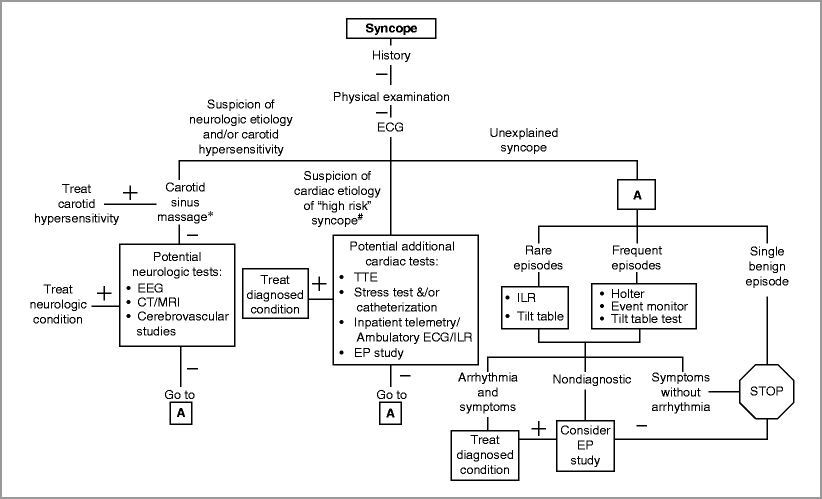
Initial Evaluation (History, Physical Examination and ECG) (Fig. 26-1)

Figure 26-1
Approach to the evaluation of syncope. Algorithm for evaluating suspected syncope. ECG electrocardiogram, ILR implantable loop recorder, Echo echocardiogram, EP electrophysiologic, SHD structural heart disease. * Contraindicated in patients with prior stroke or transient ischemic attack or with bruit present. # syncope during exercise, causing injury or motor vehicle collision or in high risk occupation (e.g. pilot)
History (To Patient and Witness If Available)
History and physical identify cause in ∼45 % of patients who are ultimately diagnosed [5]
Initial findings suggestive of organic heart disease directed additional testing leading to diagnosis in ∼8 % [5]
Helpful to differentiate between seizure and syncope (Table 26-2) [9, 10, 16]
Table 26-2
Clinical features that help differentiate arrhythmogenic syncope vs. vasovagal syncope vs. seizure
Suggestive of arrhythmia
Suggestive of vasovagal
Suggestive of seizure
Age >55
Usually younger age
Waking with a cut tongue
Duration of warning ≤5 s
Prodrome or warning symptoms
Déjà vu or jamais vu prior to loss of consciousness
Male > Female
Female > Male
Associated with emotional stress
≤2 prior episodes of syncope
History of syncope in childhood
Head turning during episode
Structural heart disease by history, exam, or echo
Diaphoresis prior to syncope
Unusual posturing or jerking limbs during episode
Exertional or supine syncope
Syncope with prolonged sitting or standing
Prolonged confusion or amnesia after episode
LBBB on ECG
Normal ECG

Stay updated, free articles. Join our Telegram channel

Full access? Get Clinical Tree


